
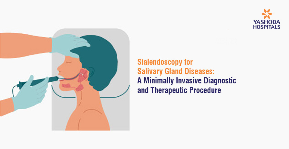
Sialendoscopy for Salivary Gland Diseases: A Minimally Invasive Diagnostic and Therapeutic Procedure
Salivary glands play an essential role in the body’s digestive system by secreting saliva into the mouth, which helps break down food and aids in swallowing. These glands can sometimes develop problems such as inflammation, obstructions, and stones, which can cause significant discomfort to patients. Salivary gland problems can occur in anyone, but they are more common in older people and those with certain medical conditions. Traditional open surgery was the only treatment option for these conditions, which is a highly invasive and risky procedure. However, advances in technology have led to the development of minimally invasive procedures such as sialendoscopy, which has revolutionised the treatment of salivary gland diseases. In this article, we will discuss what sialendoscopy is, how it works, and its benefits and risks.
What are Salivary Glands?
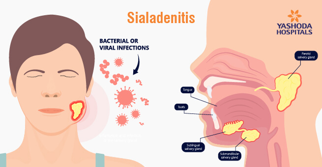
Salivary glands are exocrine glands that produce saliva in the mouth. They are classified into major and minor glands, with the major glands being the parotid, submandibular, and sublingual glands. The parotid gland is the largest salivary gland, located on the sides of the face, just above the angle of the jaw, and in front of the ears. The submandibular gland is located under the jaw, and the sublingual gland is situated below the tongue. The ducts of these glands transport saliva into the mouth, where it helps in digestion and protects the oral cavity from bacterial and fungal infections. Minor salivary glands are small exocrine glands found in the oral cavity and pharynx that secrete saliva to lubricate the mouth, aid in digestion, and protect the oral mucosa. They include labial, buccal, palatal, lingual and pharyngeal glands and differ from major salivary glands in size, location and type of saliva produced.
What is Sialendoscopy?
Sialendoscopy is a minimally invasive diagnostic and therapeutic procedure that allows physicians to visualise and treat lesions or obstructions in the salivary gland ducts. It is a highly specialised procedure that involves inserting a thin, flexible fiberoptic instrument called a sialendoscope through the mouth into the salivary ducts. The sialendoscope is equipped with a light and a video camera that allows the physician to visualise the ducts and the surrounding tissues. If a lesion or obstruction is identified, the physician can remove it using a small instrument that is attached to the sialendoscope. Sialendoscopy is performed under general or local anaesthesia, depending on the patient’s preference and the complexity of the procedure.
Sialendoscopy – Why It’s Performed
Some of the most common conditions for which sialendoscopy is recommended include:
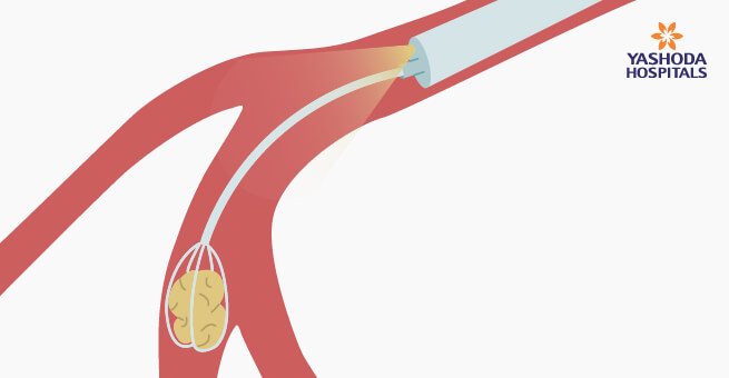
- Salivary Stones: Salivary stones, also known as sialolithiasis, are small, hard mineral deposits that form in the salivary glands or their ducts. These stones can cause pain, swelling, and infection in the affected gland. Sialendoscopy can be used to locate and remove the stone without the need for invasive surgery.
- Chronic Salivary Gland Infection: Chronic inflammation of the salivary gland, also known as sialadenitis, can cause pain, swelling, and difficulty swallowing. Sialendoscopy can help diagnose and manage sialadenitis by dilating the ducts and irrigating the gland.
- Radiation-Induced Salivary Gland Damage: Radioactive iodine treatment for thyroid gland disease can damage the salivary glands and cause inflammation, known as radioiodine sialadenitis. Sialendoscopy can help alleviate the symptoms by dilating the ducts and flushing out debris.
- Paediatric Sialadenitis: Children can develop sialadenitis due to various causes, such as viral infections, bacterial infections, or anatomical abnormalities in the salivary glands. Sialendoscopy can be used to diagnose and treat paediatric sialadenitis by identifying the underlying cause and providing appropriate treatment.
- Recurrent Salivary Gland Swelling: Recurrent swelling of the salivary glands can occur due to various reasons, such as obstructed ducts, autoimmune disorders, or tumours. Sialendoscopy can be used to examine the salivary gland and ducts, identify the underlying cause, and provide appropriate treatment.
- Sjögren’s Syndrome: Sjögren’s syndrome is an autoimmune disorder that affects the salivary glands, causing dry mouth, difficulty swallowing, and an increased risk of infection. Sialendoscopy can be used to diagnose and treat Sjögren’s syndrome by identifying any obstruction in the salivary gland ducts and providing appropriate treatment.
Advantages of Sialendoscopy
Sialendoscopy offers several advantages over traditional open salivary gland surgery.
- Minimally invasive procedure without any incisions or cuts
- Reduced pain and discomfort after the procedure
- Less scarring and a shorter recovery time
- Safer procedure with a lower risk of complications
- Small endoscope allows for precise visualisation and treatment of the affected area
- Greater accuracy and precision in removing affected tissue or stones
- More cost-effective than open surgery
- Can be performed on an outpatient basis without requiring an overnight hospital stay.
Did you know that the primary advantage of sialendoscopy is the preservation of gland functionality while reducing obstruction?
Sialendoscopy Procedure
Before the procedure, the doctor may order imaging studies such as a CT scan, MRI, or ultrasound to look at the salivary gland that is causing problems. This will help the doctor plan the procedure and identify any potential complications.
On the day of the procedure, the patient will receive either general anaesthesia or local anaesthesia with sedation. The doctor will then guide the sialendoscope into the duct of the salivary gland in the patient’s mouth. If the opening is too tight, it may be gently stretched to accommodate the scope.
Once the sialendoscope is in place, the doctor will use the light, camera, and tools to inspect the interior of the gland and remove any stones or other obstructions. The doctor may also irrigate the gland with saline to remove any debris.
After the procedure, the patient will be monitored for a short period of time to ensure that they are recovering well from the anaesthesia. Most patients can return home on the same day as the procedure.
Most patients experience little to no pain or discomfort after the procedure and can return to normal activities within a few days.
Risks of Sialendoscopy
The risks associated with sialendoscopy are generally low, but there are still some potential complications that occur in very rare cases:
- Bleeding: While bleeding is rare, there is a small risk of bleeding during or after the procedure.
- Infection: There is a risk of infection if the patient has a pre-existing condition or a weakened immune system.
- Nerve damage: There is a small risk of nerve damage during the procedure, which can cause numbness or weakness in the face.
- Perforation: There is a risk of perforation or puncture of the salivary duct or gland, which can cause further complications.
- Salivary gland damage: In rare cases, the salivary gland may become damaged during the procedure, which can lead to reduced saliva production or other issues.
In conclusion, sialendoscopy is a remarkable advancement in the field of salivary gland disorder diagnosis and treatment. This minimally invasive procedure has revolutionised the way we approach salivary gland disorders, offering patients a safe and effective alternative to traditional open surgery. By providing a targeted and precise approach, sialendoscopy has proven to be a reliable option for many patients. However, each patient’s case is unique, and consulting with a qualified doctor is the best way to determine if sialendoscopy is the right treatment option for you. So if you’re experiencing symptoms of a salivary gland disorder, don’t hesitate to speak with your doctor and explore the possibility of sialendoscopy.
References:
- Sialendoscopy
https://www.hopkinsmedicine.org/health/treatment-tests-and-therapies/sialendoscopy - Sialendoscopy: A Diagnostic and Therapeutic Aid in Salivary Gland Disorders
https://biomedpharmajournal.org/vol11no1/sialendoscopy - Sialendoscopy
https://www.baoms.org.uk/patients/procedures/38/sialendoscopy - Sialendoscopy
https://en.wikipedia.org/wiki/Sialoendoscopy


 Appointment
 Second Opinion
 Call
Call More
More


