
Brain surgery using intraoperative MRI: How does Intraoperative MRI help in neurosurgery?
What is magnetic resonance imaging (MRI)?
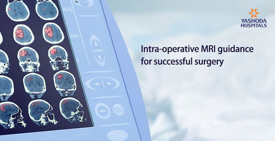
Magnetic resonance imaging is a diagnostic technique which is a safe and painless procedure to scan entire body or a body part. The MRI scanner makes use of powerful magnetic field and radio waves to provide detailed image of the scanned area. Unlike, CT scan and X-ray scan, MRI scanning does not make use of harmful radiation. MRI scans offer greater detail and accuracy over other radiological scans.
Diagnostic MRI: So far, MRI scanning is used for diagnostic purposes to identify the problem and its cause. Diagnostic MRI scans help in planning the course of treatment and surgery, if necessary.
Intraoperative MRI (iMRI/IoMRI): As discussed above, intraoperative MRI is the use of MRI scanning during the surgery. The high resolution images provided by the iMRI system lead to better precision in complicated neurosurgery operations, with the goal of reducing the risk of neurological injury during the procedure, reducing the likelihood of an incomplete resection and the eventual need for reoperation. The system also takes into account the brain shift and the movement of operating areas during the surgery, and gives real-time images, thus cutting down the chances of error drastically.
Normally, patients undergo multiple brain surgeries to cover all the procedures in a stepwise fashion. This is usually because the success of the surgery is diagnosed through regular MRI only after completion of the surgery. But with the advent of iMRI, the surgeon receives images of the surgical site immediately when the patient is on the table. This gives the surgeons and the patients an extra edge to successfully complete the surgery in one sitting. Therefore, one intraoperative MRI brain surgery equates to multiple brain surgeries without iMRI and thus substantially reduces the risks and complications for the patient.
With diagnostic MRI and intraoperative MRI combined, neurosurgeons get the real-time contrast of the diseased area that helps in reconsider marginal tumor resection, take into account the brain shift and check the progress to ensure safe and successful completion. Other than brain surgeries, iMRI is also used for other neurosurgeries, surgical oncology, radiation oncology, and ophthalmology.
What is intraoperative MRI?
Intraoperative magnetic resonance (iMRI) is the MRI diagnostic aid within the operating room for surgeries, especially brain surgeries. MRI scans help in planning the course of treatment and surgery. Intraoperative imaging process is one of the latest and cutting edge technology in neurosurgery. It offers real time picture inside the brain while performing the surgery. The image guidance it helps to minimize the risk of damaging the critical areas of the brain, identify additional resection required, and improve the chances of completing the surgery in a single sitting, successfully.
Why do we need MRI during brain surgeries?

Intra-operative MRI suite to monitor brain surgeries
Brain diseases pose a significant challenge to the neurologists and neurosurgeons. The intraoperative Magnetic resonance imaging offers following advantages:
- Real time interactive surgery: Real-time images created with iMRI are vital, as they help in accurately identifying the location of the brain abnormalities. During surgery, the brain may shift in position, thus, the location of the defect may also change. Pre-surgery images in such cases become useless. Since it enables real time correction of the nerve defects, nerve function can be evaluated during the operation.
- Accurate resection of brain tumor: iMRI helps to distinguish the margins of the tumor and normal cells and thus successful removal of an entire brain tumor. For example, brain tumours such as pediatric brain tumours, pituitary tumours and glioma. If they see remaining tumor with the iMRI, they can continue the procedure until they have removed as much of the tumor as possible, in the safest manner.
- Integrated navigation system: Integrated navigation system with real time imaging helps in navigating towards and removal of tumor or lesion in the brain. The advantage of Intra-operative mapping with electrophysiological guidance and monitoring ensures important nerve tracts and nerves are safely dealt without causing any damage. This is important in neurosurgeries that involve critical areas of brain and spine, especially in stereotactic surgeries and deep brain stimulation surgeries. Navigation and image guidance enables immediate and accurate correction as deemed necessary in the same sitting, eliminating the requirement of performing the operation again, a second time.
- No patient transportation necessary: As the patient is operated on the special table which is enabled with a sliding table top, it is simple to transfer the patient without changing the position, by just sliding the table top on to the iMRI trolley, the patient can be transferred to MRI imaging area and back to operating area. This takes care of the problems faced in transferring the patient and is also faster and economical than moving MRI models as in many hospitals.
Some of the challenges in iMRI-enabled brain surgeries are:
- Positioning of the patient:The unit has a short learning curve and adaptable easily with new design head holders available)
- Access to operating area: It is not an issue any more as MRI is in a different room and surgery can be performed freely with all independence.
What happens in the iMRI-aided brain surgery? (How it works?)
Unlike previous models of iMRI suites where the MRI was within the operating room but outside the magnetic field area, the newer models have the MRI in the adjacent room. This allows the MRI to be used as diagnostic MRI for other patients when not in use for surgery. Also it releases more space in the room and enables regular non MRI compatible instruments to be used for surgery, there by minimizing any modification of the operating room.
The patient is operated on a special neurosurgical table whose top can slide easily on to the MRI table with the patient being in the same operative posture. This helps to transfer the patient to the imaging area which is separated by a common door. Before the closure of the wound, the MRI table docks onto the special neurosurgical table top for transferring the patient. The patient is then taken for imaging with the monitors and anesthesia machines carefully in tow. Once the imaging is done the patient is then placed on the table top and slid back to the operating table. The surgery is continued as needed with the additional information obtained from the intraoperative iMRI scan.
What are the different iMRI-aided neurosurgeries?
MRI is the commonly used diagnostic tool in neurosurgery to confirm the success of the procedure before surgery is complete. The comparison of scans before and during procedure helps the neurosurgeons to decide the course of surgery forward. Hence it is useful in all neurosurgical procedures that require post-surgical evaluation, such as:
- Tumor resection for pediatric and adult brain tumors namely in intra-axial brain tumors like Gliomas, intraventricular tumors and Pituitary tumors.
- Surgery for epilepsy
- Functional neurosurgery for movement disorders like DBS
- Ablative procedures like RF lesioning
- Vascular surgeries like AVM resections,
- Skull base surgeries of tumor resections
- Endoscopic minimally invasive procedures for tumor or cysts or hematomas.
The type of surgery is decided based on the type, nature, location and severity of brain disease or damage. Some of the common brain surgeries performed are:
- Craniotomy/craniectomy is opted for tumors, aneurysm, abscess/fluid drainage, and removal of abnormal tissue. It involves an incision on the scalp and a hole (bone flap) in the skull near the area of the brain to be treated. If the hole in the skull is left open as in case of tumors, infection or brain swelling it is called as craniectomy.
- Biopsy involves removal of a small portion of brain tissue or tumor for examination under a microscope. It involves an incision on the scalp and a hole (bone flap) in the skull near the area of the brain to be treated.
- Minimally invasive endonasal endoscopic surgery involves removal of tumors or lesions through nose or sinuses without any incision. The surgeon uses an endoscope, a telescopic device with light and camera for image guidance. For example, Pituitary tumors and tumors at the base of the skull or bottom part of the brain.
- Minimally invasive neuroendoscopy surgery involves the removal of tumors or lesions through small holes in the skull.
- Ablative surgeries are also known as therapeutic brain lesioning. The process involves precisely controlled destruction of a small region of brain tissue, with help of a heat probe. This enables to improve symptoms that are produced from these brain tissues. Thalamotomy and pallidotomy are some ablative or brain lesioning procedures.
- Deep brain stimulation surgery is the surgical method which involves placing a thin metal electrode in several possible brain targets. Electrical stimulation of target brain cells improves relevant Parkinsonian symptoms.
- Image-guided stereotactic surgery also called surgical navigation, is a type of intraoperative monitoring (monitoring of the brain during surgery) that enables the neurosurgeon to precisely map the location of the tumor and determine the most effective way to remove it. The 3D image enables the neurosurgeon to see the brain during surgery to remove as much of the tumor as possible without damaging vital areas.
- Intraoperative brain mapping (awake brain surgery) brain tumor procedures while the patient is awake but sedated. This procedure is called intraoperative brain mapping, or awake brain surgery. It enables the neurosurgeons to remove tumors that would otherwise be inoperable.
What conditions require an iMRI-aided brain surgery?
Intraoperative MRI is helpful in brain surgeries that involve surgical corrections/repair, implants, biopsy, electrical stimulation aimed at resolving the problems posed by any brain tumor, cancer, disease, infection or trauma in the patient.
The brain is the centre of the body’s control and coordination in both conscious and unconscious states. However, it is an extremely delicate organ, and prone to long lasting effects from bleeding, infection, trauma and structural damage. As a result, some conditions may require brain surgery i.e neurosurgery, performed by neurosurgeons to diagnose or treat such problems.
Brain diseases present in different forms such as infections, trauma, seizures, stroke, tumors and cancers. These conditions do show signs such as swelling (inflammation), abscess, lesions, internal bleeding, clotting and vessel blockage etc. The patient may need brain surgery in the following cases:
- Structural changes: Lesions, abscesses due to infections and trauma.
- Abscess of the brain: Formation of pus within the brain due to some bacteria
- Encephalitis and meningoencephalitis: Viral induced inflammation of the brain tissues
- Meningitis: Inflammation of the lining of the brain or spinal cord attributed to infection
- Trauma: Injuries of the skull due to accidents or falls may have an impact on the brain.
- Brain cancers and tumors like gliomas, pituitary tumors, acoustic neuromas or schwannomas, medulloblastomas, lymphomas, chordomas, metastases or secondary tumours etc.

MRI surgical suite enables successful brain tumor removal surgeries
- Blood clots and bleeding: Compromised blood flow to the brain due to subdural haematoma, subarachnoid haemorrhage and intraventricular bleed etc.
- Brain aneurysm
- Cerebral oedema
- Cerebrovascular accident (CVA)
- Epidural haematoma
- Intracerebral haemorrhage (bleeding inside the brain)
- Stroke: may be ischaemic or haemorrhagic
- Subdural haematoma
- Transient ischaemic attack (TIA)
- Fluid building up in the brain: hydrocephalus
- Damage to the protective tissue called the “dura”
- Epilepsy
- Nerve damage or nerve irritation
- Parkinson’s disease
- Increased pressure after head injury
- Skull fracture
Conclusion:
The brain is the most critical part of the human body. Almost everything we do, think and speak is controlled by our brain. There are several bodily functions that the brain takes care of, even without our notice. Any condition, infection or injury affecting the brain and the nervous system can hamper the simple to complicated daily activities.
Technological advances in the field of neurosurgery have allowed high-field systems and sophistication of 3T MRI to be well integrated with the dedicated surgical suite, neuronavigation system, and digitized image transfer and projection system. This has enabled recovery from brain diseases and significant improvement in the quality of life and survival after brain surgeries.
To know more about intraoperative MRI aided brain surgeries, you can request a call back and our experts will call you and answer all your queries.
References:
- Mayo Clinic. Intraoperative magnetic resonance imaging (iMRI). Available at: https://www.mayoclinic.org/tests-procedures/intraoperative-magnetic-resonance-imaging/about/pac-20394451 Accessed on March 18, 2018
- Johns Hopkins Medicine. Neurology and Neurosurgery. Available at: https://www.hopkinsmedicine.org/neurology_neurosurgery/centers_clinics/ionm/types/intraoperative-mri.htmlAccessed on March 18, 2018
- UCLA Health. MRI Suite (Intraopperative). Available at: http://neurosurgery.ucla.edu/mri-suite-intraopperativeAccessed on March 18, 2018
- Seifert, V. “Intraoperative MRI in neurosurgery: technical overkill or the future of brain surgery?.” Neurology India3 (2003): 329.
About Author –
Dr. Anandh Balasubramaniam, Senior consultant and HOD, Neurosurgery, Yashoda Hospital, is a renowned neurosurgeon in Hyderabad. His expertise include neuro-oncology, intraoperative MRI and image guided neurosurgeries, endoscopic surgeries, endoscopic minimally invasive surgeries, deep brain stimulation and functional neurosurgeries.


 Appointment
 Second Opinion
 Call
Call More
More


