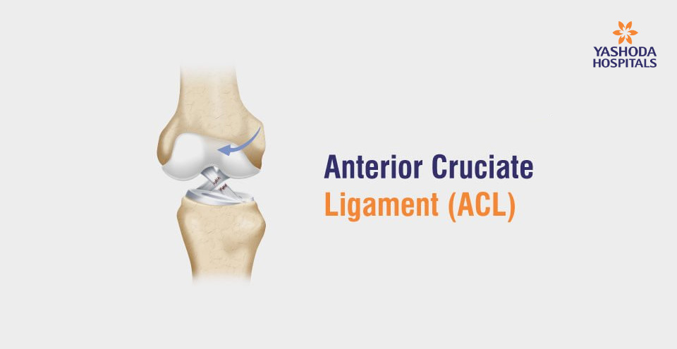
Anterior Cruciate Ligament

<< Previous Article
Spyglass CholangioscopyNext Article >>
Comprehensive Approach for Obesity వెసికోయురేటరల్ రిఫ్లక్స్ : మూత్రం వెనక్కి వెళ్లి కిడ్నీలు దెబ్బతినే పరిస్థితి!
 పీరియడ్స్ సమయంలో భరించలేని నొప్పా? అది ఎండోమెట్రియోసిస్ కావచ్చు!
 లీకీ గట్ సిండ్రోమ్ అంటే ఏమిటి? కారణాలు, లక్షణాలు, నిర్ధారణ, చికిత్స
 పంటినొప్పి : కారణాలు, లక్షణాలు, నిర్ధారణ, చికిత్స, జాగ్రత్తలు
 పాంక్రియాటిక్ క్యాన్సర్ : కారణాలు, లక్షణాలు, నిర్ధారణ, చికిత్స


 Appointment
 Second Opinion
 Call
Call More
More


