
Elastography – A promising tool for staging of breast cancer and liver diseases
Elastography is an imaging modality to assess tissue stiffness akin to palpation. The simple and intuitive relationship between palpation and elastography calls for many applications of this “palpation imaging” such as breast tumor characterization and hepatic fibrosis staging. Moreover, although palpation requires a direct contact and can only be applied to superficial organs, many elastography techniques can also be applied to deep organs opening new possibilities of diagnosis.
From a physics point of view, Elastography aims to quantitatively image the Young’s E modulus, the physical parameter corresponding to the stiffness
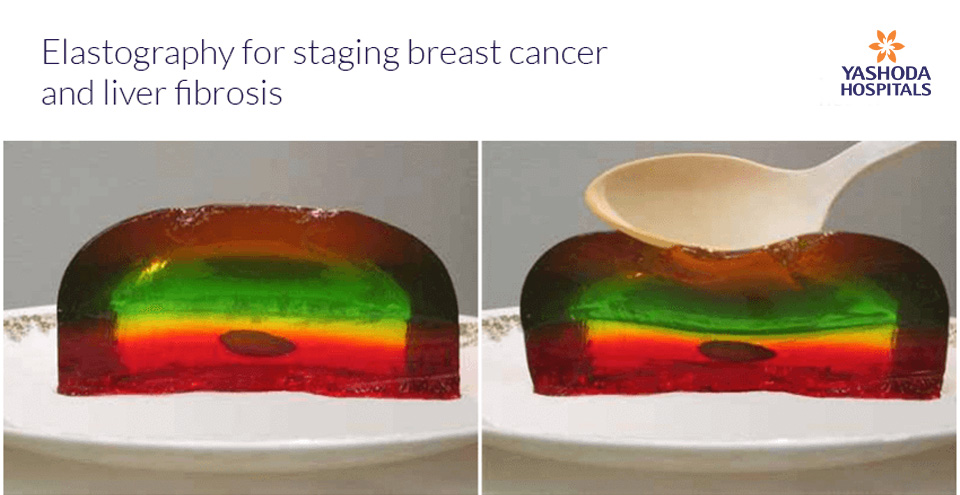
All elastography techniques rely on the same basis: an external force is applied to the studied tissue and the resulting movements are then followed on an imaging technique. Benign nodules are deformable, whereas malignant lesions tend to be stiff.

To an external compressive force, a soft lesion is more deformable, whereas, a hard lesion is less deformable relative to the surrounding tissue
Incorporation of elastography methods into ultrasound and MRI equipment has now made this modality clinically accessible.
Ultrasound Elastography:
The different methods of Ultrasound Elastography in clinical use: Quasi-static and Dynamic
- Quasi-static: Also called Strain
A constant stress is applied to the tissue. The displacement and the generated strain are then estimated using two- dimensional correlation of ultrasound images. Since the amount of stress applied is not measurable, this method is only qualitative or semiquantitative
- Dynamic method: Shear Wave
A time-varying force is applied to the tissue, either a short transient mechanical force or an oscillatory force with a fixed frequency. The most popular method is a focused ultrasound beam which generates a Shear wave or an Acoustic Radiation Force impulse in the tissue. The shear wave propagation velocity is proportional to the square root of tissue elasticity. Dynamic elastography techniques, which rely on shear waves propagation, can produce quantitative and higher resolution Young’s modulus map.
Types of Shear Wave Elastography:
- Transient Elastography one dimensional – Fibroscan. Disadvantages: Blind method with no real time visualization
- Point Shear Wave Elastography – Eg: Siemens, Phillips, This is incorporated into the ultrasound transducer, hence has the advantage of accurate placement of the ROI in the tissue
- SupersonicShear Wave Elastography – Eg: Aixplorer
 vs Shear Wave Elastography (right)")
Strain Elastography (left) vs Shear Wave Elastography (right)
Clinical applications of the two methods of Elastography
1. Quasi-static/ Strain Elastography :
Stress is applied by repeated manual compression of the ultrasound transducer. The amount of lesion deformation relative to the surrounding normal tissue is displayed as a color-coded image. It primarily helps characterize superficial structures as in thyroid, breast and the uterine cervix. A strain map helps visually interpret, with respect to a reference color bar, if the lesion is soft or hard relative to the surrounding tissue.
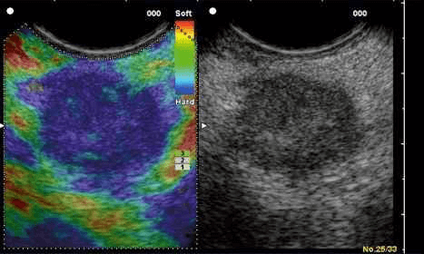
A real-time color-coded strain map (left) of the B-Mode ultrasound image (right). In this image, the tissue in blue is of hard and tissue in red is of soft consistency with the yellow-green shades representing soft-to- firm consistency, as indicated by the reference color bar to the top right of the strain image.

Strain Elastography of a firm to hard breast lesion: a fibroadenoma

Strain Elastography of a breast cyst – soft in consistency, as indicated by red on the strain map.
The disadvantages of strain imaging are that the there is significant interobserver variability and it does not quantify stiffness.
2. Dynamic / Shear Wave Elastography
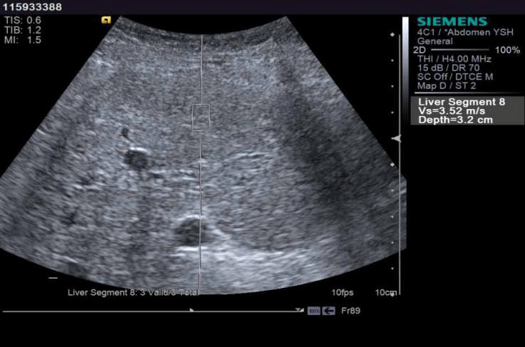
Shear wave or acoustic radiation force impulse (ARFI) from the transducer propagates in the tissue. Measurement of the propagation speed helps quantify the tissue stiffness in kilopascals or meters per second. Most important clinical use: quantitative assessment of liver stiffness. Multiple values are obtained over the right lobe of the liver and the median value is used as the index of stiffness.
Stiffer the liver, higher the recorded shear wave velocity. It is also applied in stiffness quantification in focal lesions and holds promise in stiffness imaging of spleen, kidneys and musculoskeletal system. Clinical indication for Liver Elastography:
- Detection and staging of fibrosis in patients with chronic liver disease
- Follow-up of previously diagnosed fibrosis and assessment of treatment response (Hepatitis B and C)
- Assessment for the presence of clinically significant portal hypertension (Spleen Elastography)
- Evaluation of patients with unexplained portal hypertension (Normal liver stiffness in EHPVO, NCPF). Fibrosis cutoff values for US elastographic techniques are manufacturer dependent with normal values of stiffness being below 3 m/ s or 6-7 kpa and cutoff values for Cirrhosis ranging above 2.2 m/s or 15 kpa
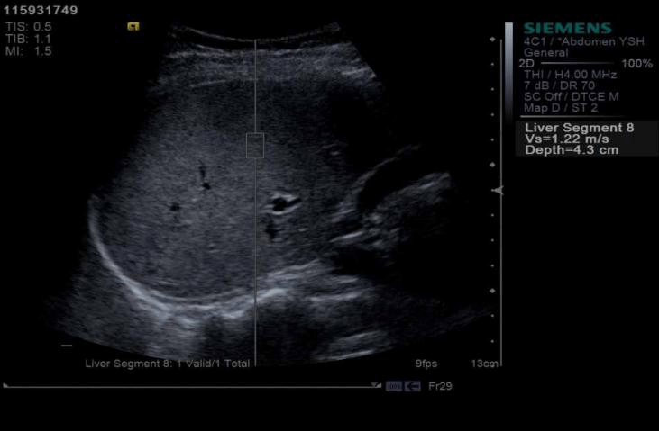

Normal liver stiffness in a young male patient with portal hypertension. Elastography helped rule out cirrhosis
Advances in Elastography
Real-time 2D Shear Wave Elastography which is now being introduced in clinical imaging allows the operator to see the generation of the elastographic measures in a color display as they are accumulated, hence helps evaluate a wider area. MR elastography incorporates the shear wave propagation generated through an external device into a wave image that displays the tissue stiffness in a color map and also quantifies the stiffness in kilopascals. Its sensitivity and negative predictive value are very high, reaching close to the gold standard of liver biopsy.
Summary
To conclude, Elastography is a novel commercially available technique for tissue stiffness imaging that helps in diagnosis and helps avoid unnecessary biopsies. In select patients, Elastography may eliminate the need for a liver biopsy for staging fibrosis.


 Appointment
 Second Opinion
 Call
Call More
More


