
A Baby Inside a Baby? The Astonishing Reality of Fetus in Fetu
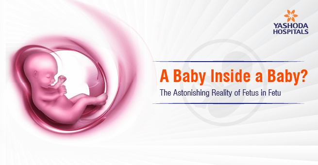
A Baby Inside a Baby? The Astonishing Reality of Fetus in Fetu
Think about this: a newborn baby looks healthy overall, but then you notice a sudden bulge in the abdominal region. Additionally, the infant experiences considerable discomfort, particularly during feeding. You take the baby to a specialist doctor, and through some tests, you discover a surprisingly rare occurrence where a fetus is living inside the body of an infant as a twin. This incident is known as “Fetus in Fetu” (FIF), a rare medical situation noticeable in 1 in 500,000 babies globally.
In this discussion, let’s gain a deeper understanding of what Fetus in Fetu (FIF) truly is, including its causes, symptoms, and methods for identification, as well as various techniques for treatment to support suffering parents and newborns.
Unraveling the Mystery of Fetus in Fetu
“Fetus in Fetu” is a rare condition in which an abnormal mass develops inside the body of a newborn. This partially formed mass can appear in unusual locations such as the skull, sacrum (the triangular bone at the base of the spine), or even inside the mouth. In some cases, it may show features like a vertebral column, limb buds, or rudimentary organs, but these are never fully developed or functional.
The expert’s thesis on how “Fetus in Fetu” occurs?
Most commonly, two theories are used for the occurrence of this rarest of rare conditions:
“Parasitic Twin Hypothesis.” In this theory, researchers estimate that “Fetus in Fetu” is an outcome of aberrant twinning, an abnormality in the typical development of a twin, which can occur due to incomplete splitting of an embryo or early fusion of two separate embryos. Another theory is the “Highly differentiated Teratoma theory,” where experts suggest that “Fetus in Fetu” is an organized and rare form of teratoma tumor. A tumour that is made up of several different kinds of tissues. These two theories focus on disturbances that occur during the embryonic development stage, leading to incomplete separation of the two embryos or early merger of the two embryos with one another.
What Does a Newborn Go Through in Fetus in Fetu?
Almost all the cases of “Fetus in Fetu” are identified in an infant newborn baby. Although some rare “Fetus in Fetu” cases have been identified in adults, sometimes. The problematic signs of “Fetus in Fetu” are most often the result of force exerted by the growing abnormal mass onto the organs of a normal newborn baby, showing signs like:
1)“Abdominal distension”, that is, abnormal swelling of the abdomen, typically this happens when the location of “Fetus in Fetu” is behind the lining of the abdominal cavity
2) “Fetus in Fetu” leads to an infant having difficulty eating, thus leading to constant vomiting due to the force exerted by the mass.
3) “Jaundice” is also a sign of “Fetus in Fetu” as there is a blockage in the functions of the liver and accumulation of bile juice.
4) “Fetus in Fetu” causes “urine retention,” that is, the accumulation of urine in the urinary tract, as there is no free movement due to the force exerted.
5) “Respiratory Problems” are caused by “Fetus in Fetu” if the mass grows in size and becomes big enough to impact the lungs.
6) “Developmental issues” are observed in a normal infant if “Fetus in Fetu” is located in the cranium and puts pressure on the brain.
7) There are a few cases of “Fetus in Fetu” where no symptoms are observed, known as “Asymptomatic cases”. These are only identified with the help of diagnostic techniques.
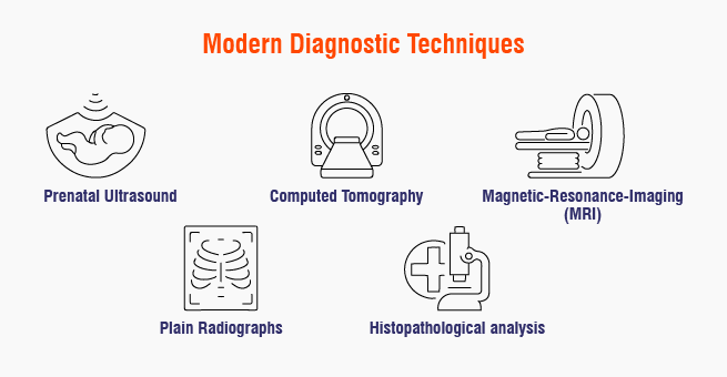
Modern Diagnostic Techniques to Detect Fetus in Fetu
Early diagnosis is very important for both treating the “Fetus in Fetu” suffering infant and his rehabilitation. The techniques listed below help achieve this;
1) A “prenatal ultrasound” often helps in finding a well-defined mass with solid bony structures inside a newborn baby.
2) “Computed Tomography (CT)” helps in getting high-resolution pictures for planning before an operation, and reveals in detail the anatomy, the presence of vertebral columns, and vascular formations.
3) “Magnetic-Resonance-Imaging (MRI)” is particularly found to be useful in the identification of “Fetus in Fetu” masses deep inside the body, besides the spinal cord.
4) “Plain Radiographs,” that is, X-rays, along with other techniques, can show Vertebral columns and bones, making sure that identified masses are “Fetus in Fetu”.
5) “Increased Serum Alpha-fetoprotein (AFP)” may be found in some cases of “Fetus in Fetu”, both in the mother and host child, starting a necessary course of action.
6) “Histopathological analysis” involves the process of tissue analysis after “Fetus in Fetu” surgery to confirm the correct diagnosis by knowing the presence of various fetal tissues.
After Diagnosis: Here’s How ‘Fetus in Fetu’ Is Treated
Treatment methods for “Fetus in Fetu” have been modernised in recent years, focusing on both the success of surgery and the overall well-being of the patient and their loved ones.
1) “Removal through surgery” is the primary method involving a curative approach, which involves complete surgical excision of the “Fetus in Fetu”. The time of surgery actually can be different, like “prenatal surgery rare”; only for cases causing serious issues detected in the uterus of the mother. “Postnatal surgery” is most commonly performed once the baby is medically stable, unless emergency intervention is needed. Great care is taken to minimize damage to surrounding structures and preserve the host’s anatomy and function. “Fetus in Fetu” mass may have its own blood supply, often from major vessels like the superior mesenteric artery or renal arteries; therefore, it is necessary to be careful during surgery.
2) “Multidisciplinary approach” for “Fetus in Fetu” is where patients benefit from coordinated, patient-centered care involving the likes of pediatric surgeons, radiologists, neonatologists, and pathologists.
3) “Postoperative management” for “Fetus in Fetu” involves intensive monitoring, which involves close monitoring for bleeding, infection, and organ function, and is essential, especially in neonates and premature infants. Family support is essential for advising parents and families, and helps them process medical and emotional challenges by reducing anxiety and promoting healing.
4) Overall progression of “Fetus in Fetu” is generally favorable; early intervention usually leads to excellent results and recovery. Rare complications can happen in some locations, especially in cranial or intra-thoracic regions, which carry increased risks and may result in severe neurological or respiratory deficit, as seen in some case reports. Recurrence & malignancy risk are minimal if the “Fetus in Fetu” is fully removed, but regular monitoring by experts is recommended.
Post Medicine: The extra therapy for “Fetus in Fetu”
If you identify a “Fetus in Fetu,” it often causes shock, confusion, and even fear among patients and families. The emotional journey can be significant, especially in cultures where medical cases often may be misunderstood or bring dishonour. Support matters as medical teams increasingly recognize the psychological toll of the “Fetus in Fetu” diagnosis, providing counseling and support resources. Sharing patient stories and increasing public knowledge helps counter the spreading of myths and develops empathy. “Fetus in Fetu” remains a subject of intense research interest, offering unique insights into embryology, twin formation, and the mechanisms behind rare congenital diseases. Ongoing investigations focus on improving prenatal diagnostic accuracy, safer surgical techniques, and long-term outcomes.
Final thoughts on the rarest of rare issues like “Fetus in Fetu”
Fetus in fetu is a rare but life-changing anomaly for those affected. Through early diagnosis, precise surgical intervention, and comprehensive care built on empathy and understanding, medical teams help children live full, healthy lives, unburdened by “Fetus in Fetu,” mass. If your child or loved one faces “Fetus in Fetu,” know that expert help is available. Recovery is not only possible, but likely; you and your family can forge ahead with renewed hope.
Have any questions or concerns about your health? We’re here to help! Call us at +918065906165 for expert advice and support.
FAQ’s
Can “Fetus in Fitu” be diagnosed before birth during the pregnancy stage?
Yes, “Fetus in Fetu” can be diagnosed by attentive specialists during prenatal sonography, often in the third trimester of the pregnancy. Though most of the cases are only identified after birth.
How rare can “Fetus in Fetu” be? And a specialist should be vigilant ?
“Fetus in Fetu” is an extremely rare condition; only about 15 cases of it have been reported in India until now. It has recently been a condition to keep a close eye on, as its identification before birth has only been possible due to the availability of modern, advanced diagnostic techniques.
What does it indicate if your ultrasound during pregnancy indicates a “Fetus in Fetu”?
This means during the early stages of pregnancy, the twin has enveloped or absorbed the other one. This then develops partially inside the normal fetus. The underdeveloped mass depends completely on the normal fetus for its nutrient supply and never develops its own vital organs.
Is surgery a safe technique for a normal baby in “Fetus in Fetu” ?
Yes, surgical removal of “Fetus in Fetu” during the delivery is completely safe medically, especially when done by expert pediatric surgeons. No long-term complications have also been observed.
Does “Fetus in Fetu” put pregnancy at risk ?
No, most of the “Fetus in Fetu” cases are carefully taken care of by transferring mothers to specialized care centres. Fetal health is carefully monitored, and any abnormal masses are removed whenever necessary. Proactive early detection of “Fetus in Fetu” most often leads to a positive outcome, with the normal fetus leading a healthy life.


 Appointment
 Second Opinion
 WhatsApp
 Call
Call More
More


