
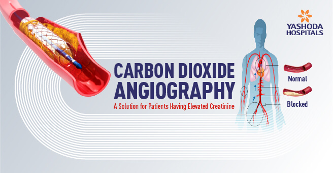
Carbon Dioxide Angiography: A Solution for Patients Having Elevated Creatinine
Contrast-induced nephropathy (CIN) is an adverse reaction to a condition when the pre-existing impairment of kidneys has taken place. Conventional angiography and angioplasty, which most often utilize iodinated contrast, may potentially injure the kidney, especially when their functioning ability has reduced.
The risk of CIN is significantly higher among patients with elevated levels of creatinine, a marker of kidney function. This severely limits treatment for these patients often when they most need it, such as angioplasty in blocked arteries, to put patients at increased risk of heart attacks and strokes. Carbon dioxide angiography offers a promising solution to this challenge. By replacing the traditional iodinated contrast agents with carbon dioxide gas, this innovative technique significantly reduces the risk of kidney injury.
Carbon Dioxide Angiography: A Game-Changer
Carbon dioxide angiography is a medical imaging technique wherein contrast agents instead of using conventional dyes based on iodine are based on carbon dioxide. It is thus very helpful for patients with kidney dysfunction because pre-existing conditions in the kidneys decline due to traditional methods of angiography. Since carbon dioxide is a natural gas, the body absorbs it very easily and exhales it out more rapidly without risking kidney function heavily. This places carbon dioxide angiography as an invaluable modality for patients with chronic kidney disease, for patients with a history of contrast-induced nephropathy (CIN), and for those in need of cardiovascular interventions without a risk of further kidney injury. Decreased CIN will positively impact the outcomes of the patients and potentially eliminate the use of dialysis or renal replacement therapy. CO2 angiography is an advanced form of vascular care.
How Does Carbon Dioxide (co2) Angiography Works?
The method uses carbon dioxide gas as a contrast medium, which is naturally occurring and well absorbed by the body and is exhaled out quickly by respiration. Once injected into the bloodstream, it displaces blood within the vascular system. Carbon dioxide has a lower X-ray absorption than blood; it produces a negative contrast effect such that when the carbon dioxide flows through the vascular system, those areas where carbon dioxide has displaced blood appear bright on the X-ray images by outlining the vascular structures and any narrowing or obstruction of the lumen.
Carbon dioxide’s lower viscosity allows for detailed visualization of intricate vascular structures in blood vessels, enabling easy diagnosis and treatment of blockages in imaging studies, especially for small blood vessels.
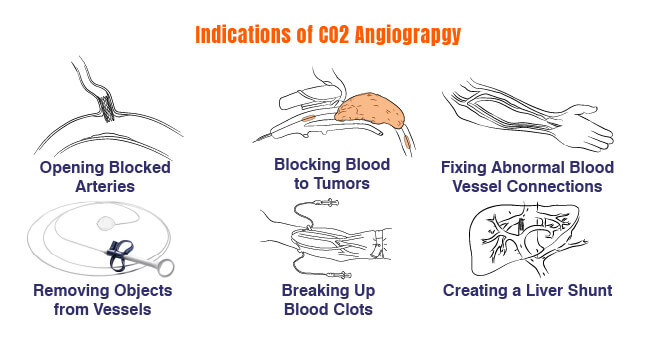
Carbon Dioxide Angiography Indications
The indications of carbon dioxide angiography involve the following:
- Angioplasty and stenting
- Tumor embolization
- Embolization of arteriovenous fistula and malformations
- Foreign body retrieval
- Catheter-directed thrombolysis
- Transjugular intrahepatic portosystemic shunt
- Transjugular liver biopsy
- Endovascular aneurysm repair (EVAR)
Carbon Dioxide Angiography Contraindications
CO2 angiography is generally safe but has certain contraindications that may pose significant risks to the patients in some exceptional scenarios:
- Absolute contraindications include thoracic aortography, coronary arteriography, and cerebral arteriography.
Rat studies show CO2 can be neurotoxic, leading to multiple ischemic infarctions and disruption of the blood-brain barrier. - CO2 should be injected into the venous limb of the graft or fistula at the anastomosis to prevent gas reflux into the brachial and subclavian arteries.
- CO2 should not be injected into the abdominal aorta in the prone position or with the patient’s head in an elevated position.
- Nitrous oxide anesthesia should be avoided as it can diffuse into the CO2 bubble, increasing CO2 volume and potentially causing pulmonary artery vapor lock.
- Relative contraindications include pulmonary hypertension and chronic obstructive pulmonary disease.
- CO2 injections should be separated by 3 to 5 minutes to prevent CO2 accumulation.
- CO2 should be used cautiously in patients with patent foramen ovale or atrial septal defect.
co2 Angiography Procedure
The process of carbon dioxide angiography primarily consists of patient selection, informed consent, positioning, catheter placement, CO2 injection, image acquisition, and image analysis. The procedure is usually used in patients having poor kidney function or allergies to iodine-based contrast agents. A patient needs to understand the risks and benefits of the procedure and obtain written consent. The catheter is inserted into an artery or vein and guided using fluoroscopy to the specific blood vessel intended for imaging. Specialized equipment is used that delivers CO2, preventing air embolism and ensuring controlled injection. Images are analyzed to determine the presence of anomalies such as blockages or narrowing of the blood vessels. The catheter is removed, and pressure is applied to stop bleeding. And finally, the patient is monitored for a short time before being discharged.
Benefits of Carbon Dioxide Angiography
Carbon dioxide angiography offers several advantages, including:
- Reduces the danger of kidney injury, which makes this safer for patients with high creatinine levels.
- Reduces patient morbidity by permitting safe cardiovascular interventions in patients with renal dysfunction
- Non-toxic contrast agent; excellent for patients with renal failure and contrast allergy
- It gets eliminated by the lungs, hence unlimited volume use, but injections have to be spaced 2-3 minutes apart.
- The viscosity of CO2 is very low; hence, it can be injected easily with a microcatheter and through guide wires.
- It is helpful in visualizing the lower extremity arteries, tumor vessels, arteriovenous fistulas, and visceral arteries.
- The CO2 reflux technique fills proximal vessels that are not visualized with contrast medium.
- CO2 is a safe and effective flushing medium for the catheter and sheath to prevent clots.
- CO2 is cheaper compared to nonionic iodinated contrast medium.
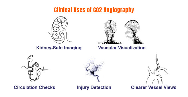
Clinical Applications
Carbon dioxide angiography has been found very promising in clinical settings in cases of:
- Chronic Kidney Disease Patients: Individuals with pre-existing kidney dysfunction undergoing coronary or peripheral angiography.
- Patients with a History of Contrast-Induced Nephropathy: Patients who have had their kidneys damaged following previous contrast procedures.
- Patients with Iodine Allergies: Some patients have severe allergic reactions to iodinated contrast media, ranging from hives and itching to potentially lethal anaphylaxis. CO2 angiography is safe for these patients.
- Vascular Interventions: CO2 is used in various vascular interventions, including stent placement, angioplasty, and aneurysm repair. It is used for visualizing vessels when deployed, ensuring proper placement for angioplasty, evaluating blood flow after angioplasty, and visualizing endoleaks after aneurysm repair, thus improving patient care.
- Peripheral Vascular Disease: CO2 angiography is used to visualize peripheral arteries and veins in patients with disorders such as atherosclerosis, peripheral arterial occlusive disease (PAOD), and deep vein thrombosis—the narrowing of the arteries and reductions in blood circulation, and the presence of blood clots.
- Trauma: CO2 angiography helps in the diagnosis and localization of internal bleeding by visualizing the source of the bleeding and assessing vascular injuries after traumatic injuries.
- Other Applications: CO2 angiography is used in a number of clinical settings, including renal artery stenosis, mesenteric ischemia, and uterine artery embolization for the treatment of uterine fibroids.
Conclusion
Carbon dioxide angiography is one of the latest innovations in vascular care. It has given a safer and more effective alternative for the patients who have elevated creatinine levels against the traditional angiography. The reduction in the risk of kidney injury allows the innovative technique to enable physicians and surgeons to intervene in a far wider group of patients who are considered inappropriate candidates for the standard procedures. As research and clinical experience continue to grow, carbon dioxide angiography is poised to play an increasingly important role in improving the outcomes of patients with cardiovascular disease and kidney dysfunction.
Yashoda Hospital’s vascular and endovascular surgeons are skilled in CO2 angiography, a new imaging technique that avoids kidney damage and allergic reactions. This technique is safer for patients with existing renal diseases or those allergic to iodine. Yashoda Hospitals uses CO2 angiography to diagnose and treat various vascular diseases, including atherosclerosis, aneurysms, and peripheral artery disease. We use minimally invasive techniques to ensure patient comfort and faster recovery times. Yashoda Hospitals is committed to providing high-quality care and is at the forefront of advanced vascular imaging and treatment options.
Have any questions or concerns about your health? We’re here to help! Call us at 918065906165 for expert advice and support.
Get Started Now To Improve Your Heart’s Health! Consult Our Specialists Right Away.
About Author –
Dr. Ranjith Kumar Anandasu
MBBS, DNB (General Surgery), MCh (Vascular and Endovascular Surgery)
About Author

Dr. Ranjith Kumar Anandasu
MBBS, DNB (General Surgery), MCh (Vascular and Endovascular Surgery)Consultant Vascular and Endovascular Surgeon


 Appointment
 Second Opinion
 Call
Call More
More


