
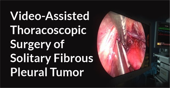
Video-Assisted Thoracoscopic Surgery Uniportal Bullectomy
Background
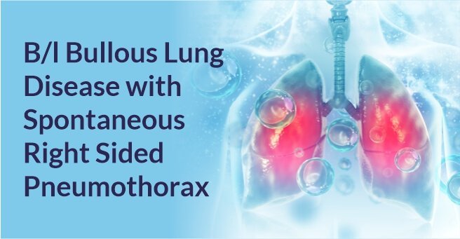
A 21 year old male patient presented with a history of dyspnea on exertion.
Diagnosis And Treatment
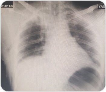
CXR showed right sided pneumothorax. ICD was placed. CT scan showed apical bullae. Uniportal bullectomy was done by Video-assisted thoracoscopic surgery (VATS).
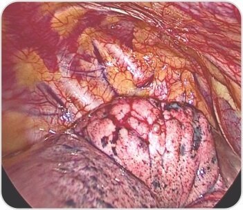
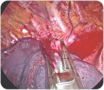
Right upper lobe apical segment bullae
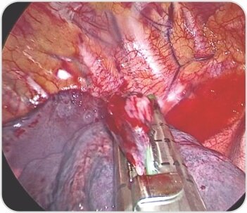
Bullectomy with endostaplers
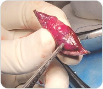
Excised bulla with portion of apical segment
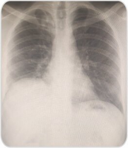
CXR at the time of discharge on POD 3
About Author –
Dr. Balasubramoniam K R, Consultant Minimally Invasive and Robotic Thoracic Surgeon, Yashoda Hospitals – Hyderabad
MS (General Surgery), MCh (CTVS)
About Author

Dr. Balasubramoniam K R
MS (General Surgery), MCh (CVTS)Consultant Robotic and Minimally Invasive Thoracic Surgeon
About Author –
Dr. Siva Prasad Goud
MBBS, DNB (CVTS) Robotic & Minimally Invasive Thoracic Surgeon
Yashoda Hospitals, Secunderabad


 Appointment
 Second Opinion
 Call
Call More
More


