
B/l Bullous Lung Disease with Spontaneous Right Sided Pneumothorax
Background
66 years old female patient presented with symptoms of shortness of breath, grade II to grade III since 2-3 months exacerbated since day 1 (grade IV). Patient is a known hypertensive and hypothyroid
Diagnosis And Treatment
On initial evaluation, X-ray was found to be radiolucent in right upper and lower zone suggestive of right sided pneumothorax. 2D Echo revealed concentric LVH / no rwma. CT chest (plain) revealed gross right pneumothorax with mild tracheal shift to left possibly due to rupture of emphysematous bullae. Subpleural emphysematous changes were noted in left lung with large bullae on both sides. Patient was taken up for emergency thoracostomy under aseptic conditions with the chest tube placed in the right 4th intercoastal space. Post procedural CXR was taken, ICD was in place. During the hospital stay, patient was closely monitored for desaturation and patient was put on oxygen support, antibiotics, nebulization, thyroid medication. Repeat CT chest revealed ICD in place with right gross pneumothorax with sub pleural bullae/ cysts projecting into pneumothorax and multiple cysts of varying sizes and para septal emphysematous bullae in left lung. Alfa 1 antitrypsin levels were normal. Patient was diagnosed as a case of B/L bullous lung disease with spontaneous right sided pneumothorax.
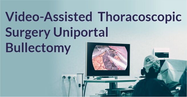
In view of the risk of recurrent pneumothorax, the patient was taken up for VATS (Video Assisted Thoracoscopic Surgery) with right sided bullectomy was done. During the hospital stay, the patient was closely monitored and the patient’s condition gradually improved and was discharged after being stabilized.
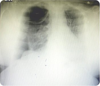
Chest radiograph showing right apical bullae
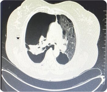
CT scan showing bullae
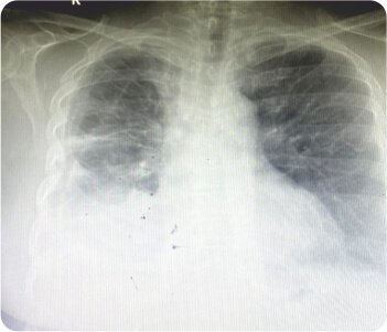
Chest radiograph at discharge
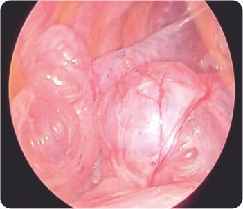
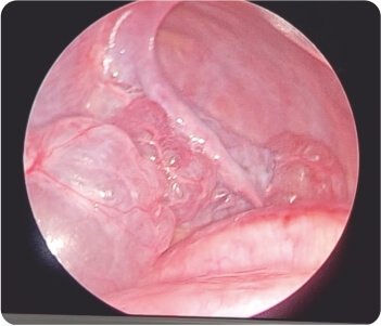
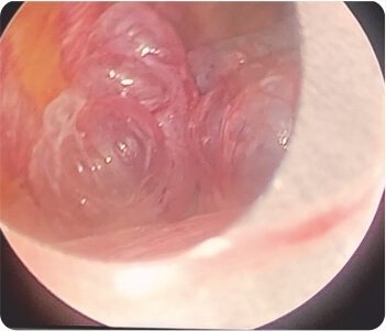
Video-assisted thoracoscopic surgery during procedure
About Author –
Dr. Balasubramoniam K R, Consultant Minimally Invasive and Robotic Thoracic Surgeon, Yashoda Hospitals – Hyderabad
MS (General Surgery), MCh (CTVS)
About Author
Dr. Balasubramoniam K R
MS (General Surgery), MCh (CVTS)Consultant Robotic and Minimally Invasive Thoracic Surgeon
About Author –
Dr. B. Vijay Kumar, Consultant Physician, Yashoda Hospital, Hyderabad
MD (General Medicine)
About Author
About Author –
Dr. Siva Prasad Goud
MBBS, DNB (CVTS)
Robotic & Minimally Invasive Thoracic Surgeon,
Yashoda Hospitals, Secunderabad


 Appointment
 Second Opinion
 Call
Call More
More


