
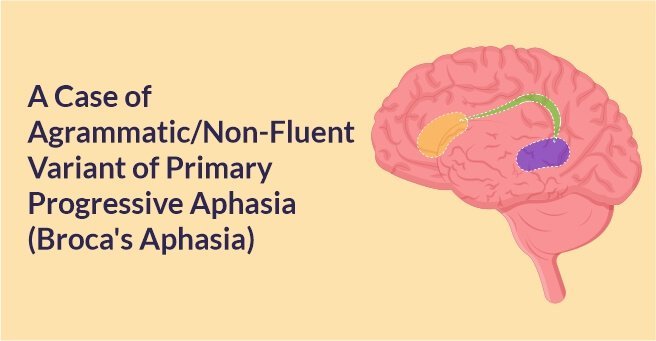
A Case of Agrammatic/Non-Fluent Variant of Primary Progressive Aphasia (Broca’s Aphasia)
Background
A 57 year old female patient presented with complaints of gradual decline in speaking ability (words per minute), speech fluency since last 4 years, associated with occasional episodes of mutism since last 6 months. She was right-handed and had the ability to write word comprehension, memory, social behaviour and visuo-spatial orientation were preserved. She had achieved a score of 29 out of 30 on mini mental scale examination (higher scores indicating better cognitive function) and score of 0 out of 3 on clinical dementia rating scale (lower scores indicating better cognitive function).
Diagnosis
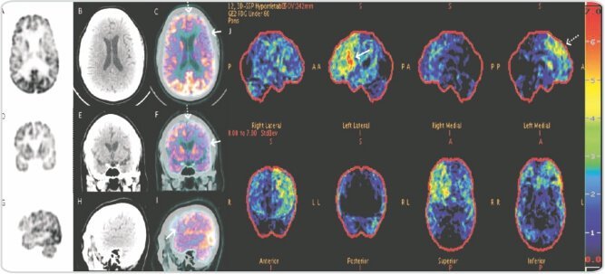
She underwent F-18 FDG PET/CT brain for further evaluation which on trans-axial (A-C), sagittal (D-F) and coronal (G-I) images revealed asymmetric hypometabolism involving the left inferior frontal cortex (solid arrow) and left anterior cingulate gyrus (dotted arrow) with no morphological abnormality on corresponding CT images. F-18 FDG PET 3D- statistical stereotactic surface projection map (J) (cortex ID, general electric software) shows asymmetric hypometabolism involving the language predominant left inferior frontal cortex (Broca’s area: solid arrow) along with left anterior cingulate gyrus (dotted arrow). Based on long time course with slow progressive deterioration of speaking ability, absence of stroke history, and selective involvement of language domain, asymmetric hypometabolism in left sided Broca’s area on F-18 FDG PET confirms a diagnosis of primary progressive aphasia.
Discussion
Compared to neurodegenerative dementia, non-fluent/agrammatic variant of primary progressive aphasia (nf-PPA) is a relatively young onset of neurodegenerative disorder characterized by language impairment and non-fluent speech with preserved memory, social, visuo-spatial and cognitive domains.
Multiple studies using magnetic resonance imaging (MRI) have reported asymmetric grey matter atrophy involving left inferior frontal cortex, adjacent perisylvian cortices with accumulation of tau in these regions on neuropathological examination (2,3). FDG PET image findings in our index case show asymmetric hypometabolism involving the similar regions suggesting synaptic failure, which precedes atrophic changes. Hypometabolism involving the left inferior frontal gyrus along with anterior cingulate gyrus in this index case suggests the involvement of frontal aslant tract, linked with the fluency of speech.
Frontal aslant tract is a recently discovered white matter tract pathway concerned with language production as documented by deep electrical stimulation brain studies (4,5). Various clinico-pathologic series and longitudinal studies reported progression of nf-PPA to frontotemporal degeneration with tau pathology (FTD-tau) and estimated conversion seen in up to 70% of patients.
Conclusion
Thus, FDG PET is proving to be a reliable imaging biomarker for early identification of nf-PPA, prediction of patients progressing to FTLD-tau and for ultimately treating tauopathies in near future.
About Author –
Dr. Kousik Vankadari
DNB, S.R (PGIMER)
Consultant & Nuclear Medicine-PET CT,
Yashoda Hospitals, Secunderabad


 Appointment
 Second Opinion
 Call
Call More
More


)