
Solitary Huge Osteochondroma of Proximal Femur in a Young Adult Without Malignant Transformation
Osteochondroma is a benign tumor containing both bone and cartilage, usually occurring near the end of the long bone. These tumors are overwhelmingly found as isolated lesions (90%) though it can present as a part of multiple hereditary exostoses.
Osteochondroma is almost certainly the result of a localized disturbance of bone growth at an Epiphysis whereby a portion of the Epiphyseal Cartilage remains in the Periosteum of the Metaphyseal Segment of the bone and then Endochondral Ossification takes place in it. Usually in continuity with the Metaphyseal Cortex and as the Cartilage Cap proliferates, Endochondral Ossification continues, resulting in growth of the Osteochondroma and with continuous bone growth the Osteochondroma moves away from the Epiphysis and tends to become hooked, its tip pointing away from the Epiphysis.
Osteochondromas grow until skeletal maturity and stop once the growth plates fuse, though slow growth from the cap may continue over time, this usually stops by age of 30.
Osteochondroma associated with multiple hereditary Exostoses may present with limb length discrepancies, increased femoral anteversion, Valgus Angulations, and Acetabular Dysplasia. Solitary Osteochondroma is most commonly asymptomatic depending on their size and location very rarely solitary Osteochondroma linked with bursal inflammation, pain, compression of neurovascular structures and malignant degeneration.
The role of proximal femoral Osteochondroma in Femoro-acetabular Impingement Syndrome and Sciatic Neuropathy very rarely reported in the orthopedic literature. Non-skeletal extrinsic complications can result from the mass effect on the adjacent tissues including muscle, tendon, nerve and vascular structures.
Nerve compression is rare and present in less than one percent of all cases of Osteochondroma.
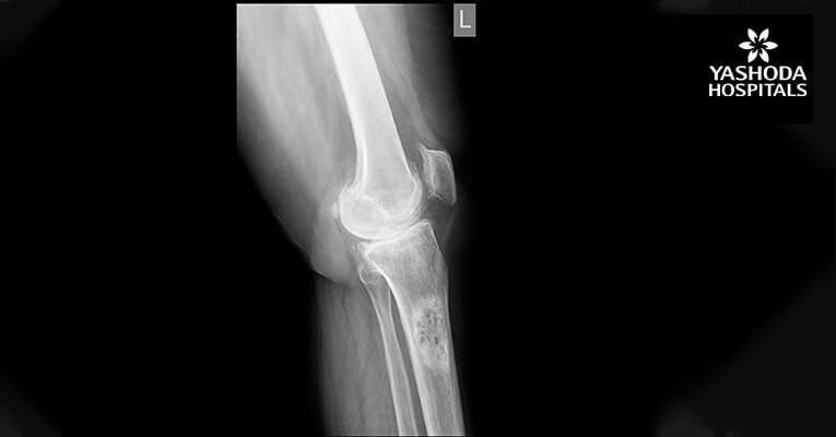
Here we report a 28 years old male present with pain in left hip and thigh due to proximal femoral Osteochondroma. Radiograph shows cauliflower-like mass originating from the left proximal femur. 3D CT scan of left thigh shows compression anterior displacement of femoral vessels near the bifurcation. This patient was treated with surgical en-bloc resection and histopathology was diagnostic of Osteochondroma.
Osteochondroma in the antero-medial aspect of subtrochanteric region of the left femur with origin abutting the lesser trochanter with a large cartilaginous cap causing compression anterior displacement of femoral vessels near the bifurcation.

pre op

Pre op X-Ray

3D CT Scan

3D CT Scan

Intra op

Intra op

Intra op

Intra op

Preop X-Ray

Post-op X-Ray
About Author –
Dr. Preshith Gaddam, Consultant Orthopaedic Surgeon, Yashoda Hospitals – Hyderabad
MS (Ortho), Fellow Sports Injuries & Ortho Trauma (NIMS), Fellow Knee & Shoulder Surgery, Arthroscopy & Sports Medicine (Germany).
His expertise includes primary hip and knee arthroplasty, revision hip and knee arthroplasty, orthopedic physiotherapy, bone trauma, correction of deformities, limb deformities & arthroscopy.


 Appointment
 Second Opinion
 Call
Call More
More


