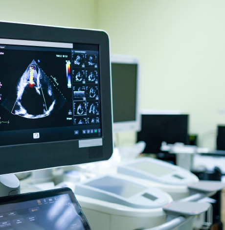
Types of 2d Echo (Echocardiography)
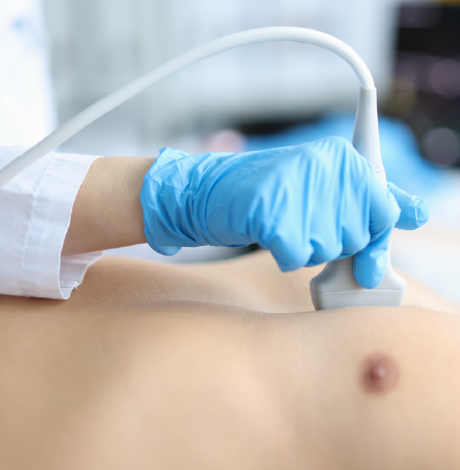
1. Transthoracic Echocardiography (TTE)
This is the most common type of 2D Echo, where the probe is placed on the chest wall to obtain images of the heart. It is used to assess heart chambers, valves, wall motion, and overall heart function in routine and emergency evaluations.
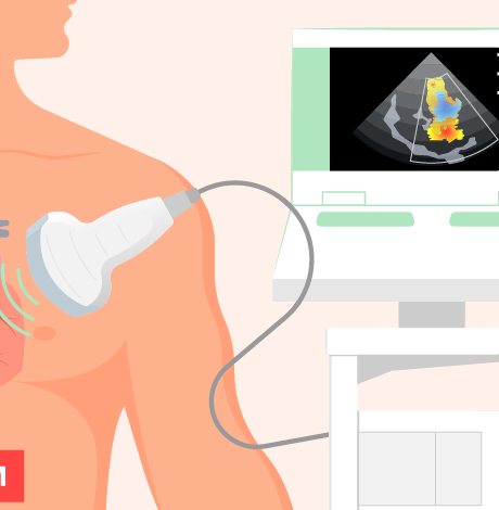
2. 2D Echo with Colour Doppler
This type combines standard 2D imaging with colour flow mapping to evaluate blood flow through the heart and valves. It helps detect valve leaks, narrowing, abnormal blood flow, and pressure changes within the heart.
3. Spectral Doppler Echocardiography
Spectral Doppler measures the speed and direction of blood flow in numerical waveforms. It is particularly useful for accurately assessing valve severity, pressure gradients, and cardiac output.
4. Stress Echocardiography
This test evaluates heart function before and after physical exercise or medication-induced stress. It is mainly used to detect coronary artery disease and assess how well the heart muscle performs under stress.
5. Transesophageal Echocardiography (TEE)
In this specialised type, a probe is passed through the food pipe (oesophagus) to obtain highly detailed images of the heart. It is used when clearer views are required, such as for detecting clots, valve infections, or prosthetic valve problems.
6. Contrast Echocardiography
A safe contrast agent is injected into a vein to improve image clarity. This helps in better visualisation of heart chambers and identification of structural abnormalities or weak heart muscle segments.
7. Paediatric / Congenital Echocardiography
This specialised echo is performed in infants and children to diagnose and monitor congenital heart defects and developmental abnormalities of the heart.
Enquire now


 Appointment
 Second Opinion
 Call
Call More
More


