Select Page

Get a Free Second Opinion

KUB Test Cost In Hyderabad

  • – Providing 30+ years of world-class healthcare to diverse communities.
  • – 600+ specialists across 62 medical specialties providing expert care
  • – Advanced technology and procedures for top-quality patient care
  • – Minimally invasive procedures for faster recovery and comfort
  • – Advanced surgical suites and ICUs for critical care
Factors Affecting

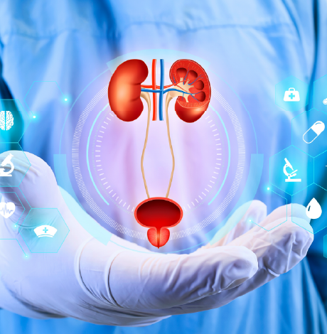
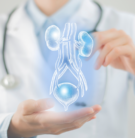
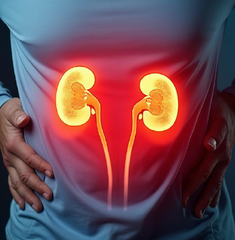
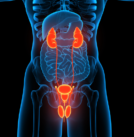
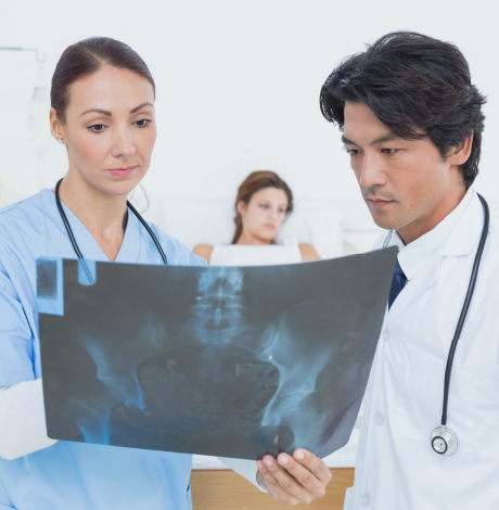
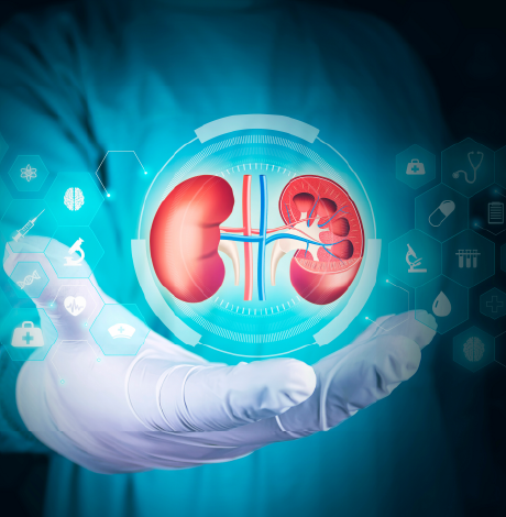
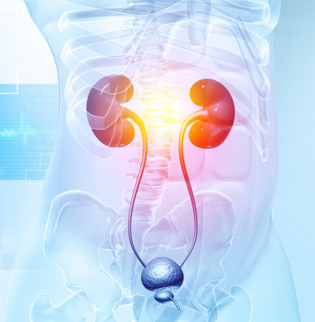
A KUB (Kidney, Ureters, Bladder) is a static image of the internal structure of the body, created by passing X-rays through the body and capturing the resulting pattern on film or a digital detector, depicting different tissues in shades of black, white, and gray on the basis of density. It is a non-invasive diagnostic imaging evaluation designed to view the urinary system and nearby abdominal organs. It is a plain abdominal X-ray evaluation that documents the interaction of ionizing radiation with internal tissues, generating images based on differences in tissue density. Broadly utilized in radiology and clinical medicine, the KUB test provides rapid insight into the anatomy of the abdomen and urinary tract, offering vital information about both normal appearances and disease-related changes. By displaying internal structures, the KUB helps clinicians correlate patient signs with underlying anatomical structures that guide clinical tests.

Unlike functional imaging methods, the KUB focuses on anatomical representation rather than physiological activity. Organs and nearby organs in the surrounding area are visible through the contrast of the radiograph, which differentiates between bone, soft tissue, gas, and calcified material. This emphasis on structures makes KUB valuable for recognising urinary tract calculi, abnormal organ outlines, displaced structures, and changed bowel gas patterns. Such results allow doctors to identify deviations from natural anatomy and to detect progression or resolution of pathological processes over time. Interpretation of KUB involves examining the density, size, shape, position, and symmetry of anatomical structures. Changes in these features indicate obstruction, inflammation, organ enlargement, calcification, and displacement. Findings are correlated with clinical history and physical evaluation to improve diagnostic accuracy and guide management.

Yashoda Hospital is a preferred choice for KUB diagnostics and treatment due to its integration of advanced imaging technology with specialized urological and nephrological expertise.

What is the cost of KUB in Hyderabad?

In Hyderabad, the cost of KUB varies depending on various factors. The range is as low as Rs 215 for basic X-ray KUB to Rs 10,000 for MRI KUB.

Factors Affecting
Factors Affecting

What are the indications for which KUB is used?

A Kidney, Ureters, and Bladder (KUB) imaging evaluation is used to test the urinary and gastrointestinal systems, whether performed by means of an X-ray, ultrasound, or computed tomography.

Primary urinary system indications include

  • Urolithiasis – Detection of stones in the kidneys, ureters, and bladder.
  • Urinary blockages – To evaluate obstructions that may cause hydronephrosis, which is swollen kidneys.
  • Hematuria – To evaluate the cause of blood in urine.
  • Infections – Evaluating problems that cause recurrent urinary tract infections or severe kidney infections like pyelonephritis.
  • Organ evaluation – Testing the size, shape, and position of the kidney and bladder
  • Tumors and cysts – Screening evaluation for abnormal masses or fluid-filled sacs in the urinary tract.
  • Congenital anomalies – Identifying defects of the urinary system at birth.
  • Post-Void Residual (PVR) – measures urine volume that remains in the bladder after urination to evaluate the functioning of the bladder.
  • Gastrointestinal and General Abdominal indications include
  • Abdominal pain – To investigate unexplained acute pain in the abdomen, flank, or back.
  • Bowel obstruction – Detecting signs of Intestinal blockage or abnormal gas patterns.
  • Gallstones – Identifying calcified stones in the gall bladder.
  • Foreign Objects – Locating swallowed items or identifying foreign bodies in the abdominal cavity.
  • Perforation – Detecting free air or fluid between abdominal organs that may indicate a ruptured stomach or intestine.
  • Procedural and Monitoring Indications Include
  • Device placement – Involves KUB confirming the correct position of stents in uretra, urinary catheters, feeding tubes, or nasogastric tubes.
  • Follow-up – KUB monitoring the progression, growth, and clearance of known kidney stones after treatment.
  • Baseline Imaging – Provides a reference image before more complex contrast studies, like an intravenous pyelogram (IVP).

Procedure of KUB

A KUB (Kidneys, Ureters, and Bladders) procedure is a diagnostic evaluation test used to examine the urinary system. Depending on your symptoms, a doctor may order an ultrasound, an X-ray, or a CT scan.

KUB Ultrasound (Non-invasive and Radiation-Free) – This is the most common first-line test, mainly for children and pregnant women.

  • Preparation involves hydration, where the patient is asked to drink approximately 1 liter of water about one hour before the scan and not urinate. A full bladder acts as an acoustic window to help the sonographer see the organs in the pelvis clearly.
  • Fasting is usually not required unless combined with a full abdominal scan.
  • The procedure is 20-30 minutes and involves the patient lying on an examination table, and a water-based gel is then applied to the abdomen and flanks.
  • A hand-held transducer is then moved over the skin to capture images in real time. The patient is asked to hold breath or change the position to give the technicians a better angle.
  • Post-Void residual (PVR) is where the patient may be asked to empty the bladder and return for a second scan to see how much urine remains.

KUB X-ray (Standard Radiograph) is used primarily to quickly identify radio-opaque kidney stones or bowel obstructions

  • Preparation is minimal, where the patient may be asked to remove jewelry or clothing with metal fasteners.
  • The procedure generally takes 5-10 minutes, where the patient lies flat in a supine position or stands against a detector. A lead apron may cover areas not being imaged for safety, and the patient may remain perfectly still and hold their breath for a few seconds while the image is captured.

Computed Tomography KUB Scan (Highly Detailed) is mostly considered a gold standard for diagnosing small kidney stones because it provides detailed 3D slices.

  • Often performed using plain (Non-contrast computed tomography) without using contrast dye, and requires minimal preparation besides removing metal. If contrast is used (Contrast-enhanced computed tomography), then 4-6 hours of fasting is required.
  • The procedure involves the patient lying down on a narrow motorized table that slides into a doughnut-shaped scanner. As the machine rotates, the patient must remain perfectly still. The patient is then asked to hold his breath for 5-10 seconds at a time.
  • It takes 10-15 minutes for a plain scan, and up to 90 minutes if contrast is involved.
Factors Affecting
Factors Affecting

Types Of KUB

There are several modalities used to perform the KUB test, which are

  • X-Ray KUB (Plain film radiography) is a common first-line diagnostic procedure that utilizes external radiation to produce images. It is commonly utilized to detect renal, uretric or stones in the bladder and evaluate organ size or position.
  • CT KUB (Computed Tomography): often utilised for more detailed imaging, a CT-KUB provides cross-sectional views of the urinary tract. A non-contrast CT KUB, as mentioned above in the procedure section, is considered the gold standard for diagnosing kidney stones.
  • Ultrasound KUB (USG KUB): is a non-invasive test that utilizes sound waves to create real-time images. It is useful for identifying kidney enlargement, cysts, tumors, and measuring post-void residual urine.
  • MRI KUB (Magnetic resonance Imaging): uses powerful magnets and radio waves to provide high-resolution images without ionizing radiation.
  • CECT KUB (Contrast-Enhanced Computed Tomography): A Computed Tomography Scan performed by using a contrast agent to better highlight the flow of blood and soft tissue abnormalities.
  • Specialized procedures are where additional tests may be ordered to evaluate specific urine functions.
  • An intravenous pyelogram (IVP) is an X-ray study that uses injected contrast dye to show how urine flows through the system.
  • A retrograde pyelogram is a procedure where contrast is injected directly into the ureters via a cystoscope.
  • Radionuclide scanning is a nuclear medicine test to evaluate kidney function and drainage.

Benefits of KUB

  • Stone detection
  • Identifying blockages
  • Structural evaluation
  • Infection and Inflammation
  • Non-invasive
  • Speed
  • Minimal preparation
  • Low or no radiation
  • Surgical guidance
  • Post-surgical monitoring
  • Incidental findings
  • Affordability
  • Wide availability

Enquire now

Factors Affecting
Factors Affecting

Factors Influencing the Cost Of KUB

  • Imaging technique used
  • Contrast dye
  • Patient factors
  • Type of facility
  • Geographical location
  • Insurance coverage

Making An Informed Decision On KUB

KUB radiography is a simple, non-invasive imaging technique used to evaluate the urinary system, particularly for diagnosing kidney stones, bowel obstruction, or abnormal gas patterns. A KUB X-ray is quick and widely available, making it useful as an initial diagnostic tool. However, it provides limited soft tissue detail and may not diagnose all abnormalities. The images require clinical co-relation and expert interpretation rather than serving as a definitive diagnosis on their own. Understanding the benefits and limitations of the KUB study helps patients and clinicians decide when it is an appropriate investigation.

Yashoda Hospital is a strong choice for KUB due to its specialized nephrology and urology institutes, advanced robotic surgeries, an experienced team for transplants, comprehensive screening packages for early detection, multispeciality team for holistic care.

Factors Affecting

Insurance Assistance for KUB treatment

  • Transparent Pricing
  • Cost Estimation
  • Billing Support
  • Insurance & TPA Assistance

Get Insurance Assistance

Free Second Opinion for KUB treatment

If you’ve been advised of the KUB treatment, get a free second opinion from our experts.

Our experienced surgeons will review your case, providing valuable insights and recommendations to help you make informed decisions about your health.

Get Free Second Opinion

Factors Affecting

Why Choose Yashoda Hospitals

Yashoda Hospitals in Hyderabad, India, is a leading option for KUB due to its top-notch team, cutting-edge technology, and comprehensive care.

blank
Comprehensive Care

Our patients trust Yashoda Hospitals for KUB treatment because its comprehensive care focuses on personalized treatment plans, minimizing complications and promoting faster recovery.

blank
Expert Doctors

Our team of experienced general surgeons delivers precise surgical care, fostering patient confidence in achieving successful outcomes.

blank
Cutting-Edge Technology

The Centre for General Surgery at Yashoda Hospitals offers state-of-the-art facilities and the latest technologies.

blank
Clinical Excellence

Yashoda Hospitals is recognised as the Best Hospital for KUB treatment Cost In India , offering exceptional surgical care services.

All major insurance are accepted for
KUB Test Cost In Hyderabad Treatment


Our Locations

  • Malakpet Location

    Malakpet

  • Somajiguda Location

    Somajiguda

  • Secunderabad Location

    Secunderabad

  • Hitec City Location

    Hitec City

FAQ's

Yes, emerging explainable AI (XAI) models, such as those utilising VGG16 architecture, have achieved upto 97.41% accuracy in identifying kidney stones that may be challenging for human eyes to diagnose standard KUB X-rays.

It is becoming a preferred trend for evaluating complex cystic renal lesions. CEUS uses microbubbles that are non-nephrotoxic, making it safer than traditional computed tomography contrast for patients with significant kidney failure.

Yes, because it covers the lower abdomen, a KUB ultrasound can incidentally detect appendicitis, ovarian cysts, or even pelvic masses that were not the original reason for the scan.

This rare but important measurement utilizes Doppler ultrasound to calculate a ratio of blood flow velocity. A difference of more than 0.1 between kidneys is a highly sensitive indicator for an acute ureteral obstruction, often utilized in pregnant patients to avoid radiation.

No, even though KUB is considered a safe test during pregnancy. It is specifically calibrated to look at the mother's urinary system and is not intended for prenatal fetal monitoring.

Terms and conditions

All the cost and surgery information provided on the website is primarily to enable the users to obtain a better understanding of Yashoda Hospitals and the medical services provided by it, and the information thereby provided is subject to changes and modifications as and when required without any prior notice. Actual cost of the surgery details would be confirmed at the hospital after doctor consultation only.

Yashoda Hospitals does not represent or endorse the accuracy, completeness or reliability of any information displayed, uploaded or distributed through the website. The usage of the content implies that you acknowledge that any reliance upon any such statement or information shall be at your own risk. Yashoda Hospitals does not resume any responsibility related to the accuracy of information provided or made available. None of the individual developers, system operators, third-party contributors and management of Yashoda Hospitals or anyone else connected to Yashoda Hospitals can be held responsible for the results or consequences incurred due to any reliance being placed or adopt any of the information presented on this website.