Select Page

Get a Free Second Opinion

EEG Test Cost In Hyderabad

  • Providing 30+ years of world-class healthcare to diverse communities.
  • 600+ specialists across 62 medical specialties providing expert care
  • Advanced technology and procedures for top-quality patient care
  • Minimally invasive procedures for faster recovery and comfort
  • Advanced surgical suites and ICUs for critical care

An electroencephalogram is a non-invasive identification and testing device utilized to document and test the electrical activity of the brain by identifying and magnifying signals created by neuronal activity in the cerebral cortex region of the brain. Vastly utilized in neurology and neuroscience, the electroencephalogram allows an instant chance of observing the functioning of the brain and gives very important knowledge of both normal physiological processes and disease conditions. An electroencephalogram shows the brain’s actions as wave patterns that demonstrate the common functioning of a large number of neurons, thereby concentrating on functional tasks instead of brain structure. This makes it very useful in studying the brain’s rhythm, synchronization of neurons and any changes in consciousness, like being alert, relaxed or in sleep. An electroencephalogram documents the electrical activity of cortical pyramid neurons by measuring the excitatory and inhibitory postsynaptic potentials produced by synchronized neuronal firing. Brain performance is visualized by waveforms specified by frequency, amplitude, shape and symmetry. These change dynamically based on external stimulus or internal conditions.

Electroencephalogram results are obtained by using scalp electrodes placed according to standardized systems like the International 10-20 system. A key advantage of the electroencephalogram is its high temporal resolution, allowing for the identification of any rapid change in neurons. Although its spatial resolution is minimal electroencephalogram is still highly useful for clinical diagnosis and research.

What is the cost of an EEG in Hyderabad?

Electroencephalogram evaluations are available in several forms based on the method and the length of the brain’s activity recording. The cost of an electroencephalogram varies based on the type of test and the diagnostic facility. The approximate cost of an electroencephalogram in India and Hyderabad is mentioned below.

Factors Affecting
Factors Affecting

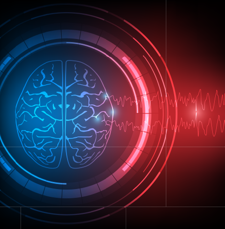
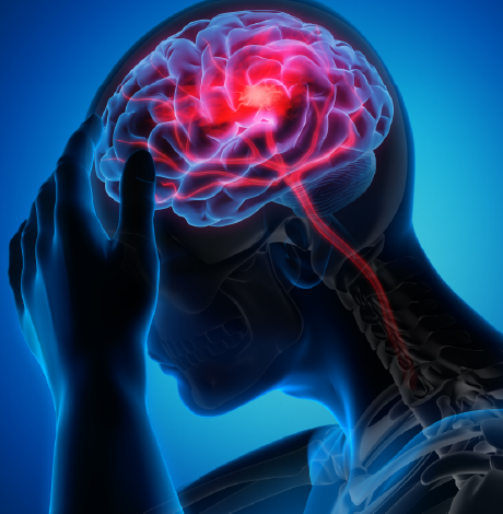
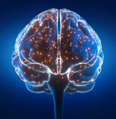
Indications of electroencephalogram (EEG)

An electroencephalogram is utilized every time there is a requirement to examine the functional electrical activity in the brain. It is mainly utilized in conditions that involve seizures, altered consciousness, diffuse cerebral dysfunction, and unusual cortical activity.

Epilepsy and seizure disorders:
The major indications of an electroencephalogram include epilepsy and seizure disorders. An electroencephalogram is useful in confirming the presence of epileptic activity by diagnosing unusual electrical discharges in the brain. It helps in differentiating epileptic seizures from non-epileptic scenarios. Whether it is the classification of seizures or identifying if seizures start from a focal area or involve the complete brain electroencephalogram, it helps in keeping track of antiepileptic therapy and examining patients with repeated or unexplained seizure occurrences.

Altered level of consciousness:
Electroencephalogram is also used in patients with sudden changes in conscious states, namely, confusion, delirium, stupor or coma. It helps in evaluating the overall degree of cerebral dysfunction and provides information about brain activity in patients who do not usually respond. Electroencephalogram patterns can detect the depth of unconsciousness and help distinguish between metabolic causes or damage in the brain structure.

Encephalopathies:
An electroencephalogram is widely used in the examination of encephalopathies, which are widespread disorders of brain function. They may be caused by unusual metabolism, toxins, hypoxia, or systemic illness. An electroencephalogram depicts the declining functioning of the brain in such conditions. It is useful in evaluating severity, tracking progression, and examining the response to treatment of metabolic, toxic, hypoxic, hepatic and uremic encephalopathies.

Brain infections:
An electroencephalogram is also used in suspected infections of the brain. Namely, encephalitis and meningoencephalitis. It helps in diagnosing abnormal cortical activity caused by either inflammation or infection. Certain electroencephalogram patterns help in early diagnosis and provide information about the extent and localization of brain involvement, especially when clinical results are not clear.

Head injury and Traumatic brain injury :
An electroencephalogram is useful in assessing the functioning of the brain after injury to the brain. It helps in diagnosing diffuse or focal problems in electrical activity. An electroencephalogram is also used on patients with post-traumatic seizures or those who suffer from neurological problems after trauma.

Stroke and cardiovascular problems:
Electroencephalogram is utilized in patients with stroke to evaluate the involvement of of cortex and how far its functions have been affected by stroke or cardiovascular issues. It may lead to unusual electrical activity in the brain. An electroencephalogram also keeps a track of recovery and helps identify problems like seizures that may occur after a stroke.

Brain tumours and space-occupying lesions:
An electroencephalogram helps in the detection of focal abnormalities in brain’s electrical activity caused by cortical irritation or compression. This also provides information about how lesions affect the cortical function and electroencephalogram, then finds its use as a supportive tool for imaging studies.

Sleep disorders:
In case of sleep disorders electroencephalogram documents the brain’s activity during different stages of sleep. This helps in the identification of unusual sleeping patterns and sleep-related seizures. An electroencephalogram is also used as a key component in polysomnography.

Dementia and neurodegenerative problems:
Electroencephalography evaluates cortical dysfunction in patients with reduced cognitive ability. Shows a gradual decline in the brain’s activity in advanced stages. An electroencephalogram helps in clinical diagnosis and tracking disease progression in cases of dementia or other neurodegenerative disease conditions.

Developmental and pediatric neurological disorders: Helps in evaluating the brain’s activity in infants and children who are facing a delay in their development. Along with that, it helps in detecting neonatal seizures and childhood symptoms of epilepsy. Also evaluates brain maturation and unusual electrical patterns.

Behavioural and psychiatric conditions:
Electroencephalography identifies neurological reasons for behavioural changes and helps in differentiating events of epilepsy from psychogenic disorders. Also supports testing of unexplained mental and behavioural symptoms.

Monitoring the functioning of the brain in critical care:
In these patients, the electroencephalogram continuously keeps a track of seriously sick cases, and helps in diagnosing ongoing or non-convulsive seizure activity. It also evaluates changes in cerebral functioning over time.

Monitoring during Anesthesia and Surgery, the electroencephalogram records the depth of anesthesia during a surgical procedure and evaluates cerebral activity during major operations. Also helps in detecting cerebral ischemia during the operative procedure.

Brain death examination:
An electroencephalogram helps in identifying the absence of cerebral electrical activity and supports clinical identification of brain death, utilizing it in combination with other tests of confirmation.

Unexplained neurological symptoms:
Examines unexplained loss of consciousness or fainting, and evaluates abnormal movements or sensory disturbances. Also helps in the identification of any unusual brain activity.

Enquire now

Procedure Of Electroencephalogram:

The electroencephalogram procedure is a non-invasive technique used to document the electrical activity of the brain through electrodes placed on the scalp. It is performed under controlled conditions to get precise and authentic brain wave recordings while ensuring sure of patient’s comfort and safety. The procedure involves systemic preparation, placement of the electrode, recording and documentation.

Pre-procedure preparation:
Before the beginning of the electroencephalogram procedure, attention to certain basic steps is required to ensure precise recording and the comfort of the patient. Identification of the patient is ascertained, and the objective of the electroencephalogram test is clarified. Useful medical history, neurological symptoms, and medicines that the patient is on are reviewed. The scalp is then evaluated for cleanliness, wounds or infections of the skin. The hair of the patient should be clean and dry to allow proper contact of the electrode.

Patient Positioning:
Proper positioning of the patient is essential to obtain clear electroencephalogram recordings. The patient is requested to lie down on a bed or sit comfortably in a reclining chair. The head and the body are supported to promote relaxation. The patient is then asked to remain still and relaxed during an electroencephalogram recording.

Placement of electrode:
Placement of the electrode is an important step as electroencephalogram recordings depend on the precise identification of electrical signals from the scalp. Standard scalp landmarks are identified using anatomical reference points. Electrodes are arranged according to the 10-20 international system. A conductive gel or paste is applied to decrease the electrical resistance. The electrodes are then firmly secured to avoid any movement during recording.

Set up of equipment and calibration:
The electroencephalogram equipment must be properly connected and checked to ensure reliable documentation of signals. Electrodes are connected to the electroencephalogram amplifier and recording system. Electrode impedance is then evaluated to confirm good electrical contact. The encephalogram equipment is then calibrated before the start of recording.

The recording phase of the electroencephalogram:
This is the exact phase where the electrical activity of the brain is recorded by an electroencephalogram. Continuous brain activity is recorded through the electrodes on the scalp. Patient is advised to relax and remain still throughout the evaluation, and is instructed to open eyes and close eyes at regular intervals. Standard activation techniques may be performed, such as hyperventilation (rapid and deep breathing) and photic stimulation using flashing lights.

A routine recording of an electroencephalogram usually takes 20 to 40 minutes.

Observation during recording:
Continuous observation is vital to correlate the results of electroencephalogram findings with patient behaviour. The technician closely observes the patient for movements or symptoms. Any clinical events, such as unusual movements or altered awareness, are noted. Artifacts caused by blinking, muscle activity or movement are identified and minimized.

Completion of procedure:
Once sufficient data is recorded, the electroencephalogram procedure is concluded safely. The recording is stopped after the required information is gathered. Electrodes are then removed from the scalp, followed by the conductive gel being removed from the patient’s hair or skin.

Post-procedure care after electroencephalogram:
After an electroencephalogram procedure, the patient requires minimal care and can start normal activities. The patient is reassured and allowed to leave the testing area. No recovery period or restriction is necessary. Electroencephalogram recordings are saved for detailed evaluation.

Documentation or reporting of the electroencephalogram:
Proper documentation ensures accurate interpretation and future reference. Patient details and test conditions are recorded. Duration and type of encephalogram performed are documented. Any unusual findings or events during the procedure are noted. The Electroencephalogram is then reviewed and interpreted by a trained specialist.

Factors Affecting
Factors Affecting

Types of EEG

In 2025, electroencephalograms are classified by their clinical procedures, the technology used for recording, and the specific brain activity they aim to capture.

Clinical electroencephalogram tests:

These are standard tests used in clinics and hospitals to identify epilepsy and sleep disorders. The tests are mainly.

Routine electroencephalogram tests are used to diagnose seizures and general brain dysfunction.

Sleep-deprived electroencephalogram: is conducted after a patient has stayed up all night for part or all of the previous night to trigger abnormal activity that may not appear when fully rested.

Sleep electroencephalogram (Polysymnogram): is conducted when the patient is sleeping, and is mostly used to identify nocturnal epilepsy or sleep disorders like sleep apnea.

Ambulatory electroencephalogram: is a portable examination where the patient wears a small recorder for 24 to 72 hours while they continue with normal activities.

Video electroencephalogram (Video telemetry): Combines electroencephalogram recording with synchronized video of the patient to match physical movements with specific brain wave patterns.

Prolonged electroencephalogram: is a longer version of the routine electroencephalogram, often lasting over an hour, to capture infrequent signs.

Invasive (Intracranial) electroencephalogram: is a surgical procedure where electrodes are placed directly on the brain’s surface (electrocorticography) or inside the brain (stereo-EEG) to pinpoint origins of seizure before surgery.

Continuous electroencephalogram (cEEG): used in ICUs for continuous monitoring of critically ill or unconscious patients.

Specialized and Emerging Electroencephalogram Types:

This includes Evoked potential studies: measure the brain’s electrical response to specific sensory stimuli like flashes of light or sounds.

Quantitative electroencephalogram (qEEG): uses computer algorithms to evaluate EEG data into a map of the brain, often utilized in research or neurofeedback.

24/7 Subcutaneous electroencephalogram (SubQ): A newer technology involving a long-term electrode implanted under the skin for up to 15 months to monitor treatment-resistant epilepsy.

Event-related potentials (ERP) are a research-focused electroencephalogram type that records the brain’s response to specific cognitive or sensory events.

Technology-based Electroencephalogram:

This includes Digital electroencephalogram: is a modern system that converts brain signals into digital data for advanced computer analysis.

High-density electroencephalogram: uses a large number of electrodes to accurately map the brain activity.

Wireless or Wearable electroencephalogram: Headsets and portable devices allow for movement and brain-computer interface applications.

Dry vs wet electrode encephalogram: Wet electrodes utilize conductive gel for higher signal quality. While dry electrodes use a special material for a faster or cleaner setup.

Factors Affecting
Factors Affecting

By Brain Wave frequency:

This includes Delta (0.5 to 4 Hz), which is represented by deep sleep,
Theta (4-8 Hz) is associated with drowsiness and light sleep,
Alpha (8-13 Hz) represents relaxed wakefulness with eyes closed,
Beta (13-30 Hz) is associated with active thinking and alertness, and
Gamma (above 30 Hz) is linked to high-level cognitive tasks and memory.

Side Effects of Electroencephalogram:

An electroencephalogram is a non-invasive, painless method that generally has no side effects. The test records brain activity rather than stimulating it, so there is no risk of an electric shock. Potential temporary side effects include skin irritation, lightheadedness or tingling, fatigue, and sticky residue. Risks due to the electroencephalogram that are linked with specific conditions include triggering of seizures and sedation effects.

Benefits of the Electroencephalogram

  • Diagnosis of Epilepsy and Seizure Disorders
  • Sleep Disorders
  • Brain Injuries and Diseases
  • Cognitive and Mental Health
  • Critical Care Monitoring
  • Non-invasive Safe
  • Real-time Data
  • Cost Effectiveness
  • Poratbility
  • No recovery time

Factors Influencing The Cost of Electroencephalogram

  • Type and Duration Of Test
  • Labour and Expertise
  • Technology and Equipment
  • Facility and Operational Factors
  • Geographic and Insurance Factors

Enquire now

Factors Affecting
Factors Affecting

Making An Informed Decision On Electroencephalography

Electroencephalography is a non-invasive technique for measuring the brain’s electrical activity and is widely utilized for studying seizures and sleep disorders. While an Electroencephalogram offers excellent time resolution, it has a limited ability to accurately locate the brain’s activity. Electroencephalogram results show patterns that require expert interpretation rather than direct diagnosis. Knowing its benefits and limits helps patients make an informed decision about using an electroencephalogram.

Yashoda Hospitals is a preferred choice for an electroencephalogram primarily due to its advanced neuroscience institute and specialized epilepsy centre. Which offer complete diagnostic and treatment services.

Why Choose Yashoda Hospitals

Yashoda Hospitals is committed to providing world-class treatment for patients from across the globe. With the unique combination of state-of-the-art technology, intuitive care, and clinical excellence, we are the healthcare destination for thousands of international patients in India.

blank
Comprehensive Care

On the journey to good health, we understand that it is important for you to feel at home. We plan out all aspects of your trip.

blank
Expert Doctors

Experienced specialists perform non-invasive and minimally invasive surgeries to provide the best treatment for international patients.

blank
Cutting-Edge Technology

Our hospitals are equipped with advanced technology to perform a wide range of procedures and treatments.

blank
Clinical Excellence

We deliver excellence by delivering quick and efficient healthcare and through pioneering research that helps all our future.patients.

All major insurance are accepted for
EEG Test Cost In Hyderabad Treatment


Our Locations

  • Malakpet Location

    Malakpet

  • Somajiguda Location

    Somajiguda

  • Secunderabad Location

    Secunderabad

  • Hitec City Location

    Hitec City

FAQ's

An electroencephalogram can diagnose a brainwave pattern linked to attention or relaxation. But it cannot directly measure emotions.

Yes, an electroencephalogram is non-invasive and safe for repeated or long-term monitoring, with no known harmful effects.

Clinically electroencephalogram is medically regulated and diagnostic, while a wearable electroencephalogram is designed for wellness, training or self-tracking -not diagnosis.

Consumer electroencephalogram devices provide basic brainwave data but lack the accuracy, electrode coverage, and reliability of medical-grade electroencephalogram devices.

Knowing your purpose and electroencephalogram strengths and limits helps you make an informed decision.

No, an electroencephalogram is not painful; it is a non-invasive technique that only measures the activity of the brain utilizing scalp electrodes and does not give any electric stimulus.

An electroencephalogram cannot directly diagnose a brain tumour because it does not show the brain’s structure. However, it may show unusual brain activity that shows a tumour is affecting the brain function.

A routine recording of an electroencephalogram usually takes 20 to 40 minutes. However, the range may extend to several days depending on the type.

Terms and conditions

All the cost and surgery information provided on the website is primarily to enable the users to obtain a better understanding of Yashoda Hospitals and the medical services provided by it, and the information thereby provided is subject to changes and modifications as and when required without any prior notice. Actual cost of the surgery details would be confirmed at the hospital after doctor consultation only.

Yashoda Hospitals does not represent or endorse the accuracy, completeness or reliability of any information displayed, uploaded or distributed through the website. The usage of the content implies that you acknowledge that any reliance upon any such statement or information shall be at your own risk. Yashoda Hospitals does not resume any responsibility related to the accuracy of information provided or made available. None of the individual developers, system operators, third-party contributors and management of Yashoda Hospitals or anyone else connected to Yashoda Hospitals can be held responsible for the results or consequences incurred due to any reliance being placed or adopt any of the information presented on this website.