
2D-Echo Test
The 2D Echocardiography (2D Echo) test is a non-invasive imaging method used to examine the heart’s structure and function in real time. It uses high-frequency sound waves (ultrasound) to create detailed, moving images of the heart chambers, valves, and surrounding structures. The 2D Echocardiography test provides valuable information on how successfully the heart pumps the blood and whether there are any abnormalities in its structure or function. It is broadly used in the diagnosis and monitoring of various heart issues that include valve diseases, heart failure, congenital heart diseases, and cardiomyopathies. Since the procedure is safe, painless, and does not expose the patient to radiation, the 2D Echo test has become an important tool in modern cardiology for both initial examination and ongoing patient care.
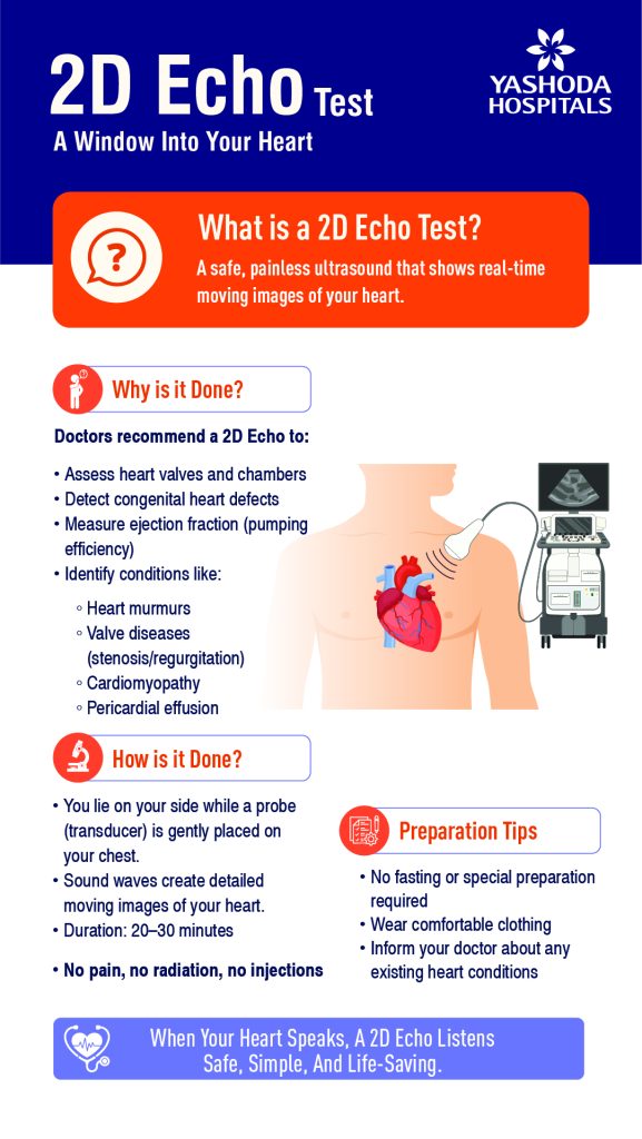
Uses of the 2D Echo Test:
- The 2D Echo test is utilized to examine the structure and function of the heart. It assists in identifying heart valve diseases such as stenosis and regurgitation.
- The 2D Echo test is useful in identifying heart failure and evaluating the pumping efficiency of the heart (ejection fraction).
- It helps in the identification of congenital heart defects, along with assisting in identifying fluid accumulation around the heart (pericardial effusion).
- The 2D Echo test is also used to monitor the progression of heart diseases and the response to treatment given for heart diseases.
Indications of the 2D Echo Test:
- The 2D echo test is suggested for patients who complain of chest pain or shortness of breath.
- It is also suggested when there are symptoms like palpitations or irregular heartbeat.
- The 2D echo test is recommended in patients with a history of heart attack or heart failure, along with patients whose ECG or chest X-ray shows abnormal findings.
- The 2D Echo test is also suggested for examining patients with congenital heart abnormalities.
Preparation For 2D Echo Test:
- The patient needs no special preparation, or practice fasting before undergoing a 2D Echo evaluation.
- The patient should wear comfortable clothing for easy access to the chest area. The patient should inform the doctor about them dealing with any previous heart issues or taking medicines.
- Doctors and healthcare professionals advise patients to remain calm and relaxed before the start of the 2D Echo test.
Procedure Of 2D Echo Test:
- The patient is advised by a doctor or healthcare professional to lie down on the evaluation table, normally on the left side.
- A special gel is applied to the chest area to improve the transmission of the sound wave. Followed by a device called a transducer, which is placed on the chest, that emits ultrasound waves.
- These sound waves reflect off the heart structures and create real-time images on the monitor. The technician then might move the transducer to obtain images from different angles.
- The 2D Echo test procedure is painless and normally takes about 20 to 40 minutes to complete.
Benefits of the 2D Echo Test:
- The 2D Echo test is a non-invasive and completely safe procedure, with no exposure to any radiation.
- It provides real-time visuals of the heart’s function and structure. The test helps in the early identification of heart diseases.
- The 2D Echo test is useful for monitoring treatment progress in cardiac patients, as the procedure is fast, painless, and widely available.
Factors Influencing 2D Echo Results:
- Poor image quality due to obesity or lung disease might affect the accuracy.
- If the patient is not still and moves during the test, this can interfere with image clarity.
- The experience and technique of the 2D Echo device operator can also impact/Influence outcomes.
- Some of the heart conditions might need the use of additional imaging techniques for confirmation.
Types of 2D Echocardiography:
The Transthoracic Echocardiography (TTE) is the most common type of 2D Echocardiography, where images are obtained by placing the transducer on the chest wall. It is straightforward, non-invasive, and widely used for routine examination.
The Transesophageal Echocardiography (TEE) involves thrusting a probe into the esophagus to obtain more detailed pictures of the heart. This gives a clear visualization, mainly of posterior heart structures, but it is slightly more invasive.
The Stress Echocardiography is a type of 2D Echocardiography that is performed while the heart is under physical stress, either due to exercise or medications. It assists in examining how the heart functions during increased workload and is useful in identifying coronary artery disease.
The Doppler Echocardiography especially examines blood flow through the heart and assists in diagnosing abnormalities such as valve leaks or blockages.
Interpretation Of 2D Echocardiography Results:
- A normal ejection fraction (EF) ranges from 55% to 70%, suggesting that the heart is pumping successfully.
- An ejection fraction (EF) below 40% might indicate heart muscle failure or weakened heart muscle.
- Normal heart chamber sizes and valve function indicate a healthy heart. Abnormal findings might include enlarged heart chambers, defects in the valves, or reduced pumping ability of the heart.
- The presence of fluid around the heart or abnormal blood flow patterns might indicate specific cardiac conditions.
- The Interpretation of 2D Echocardiography should always be done by a cardiologist in correlation with other clinical findings.
Side Effects Of 2D Echocardiography Test:
- The 2D Echocardiography test is usually very safe and does not have any significant side effects.
- The patient might feel slight discomfort from the pressure of the transducer on the chest.
- The gel applied might feel cold, but it does not cause any harm.


 Appointment
 Second Opinion
 Call
Call More
More


