


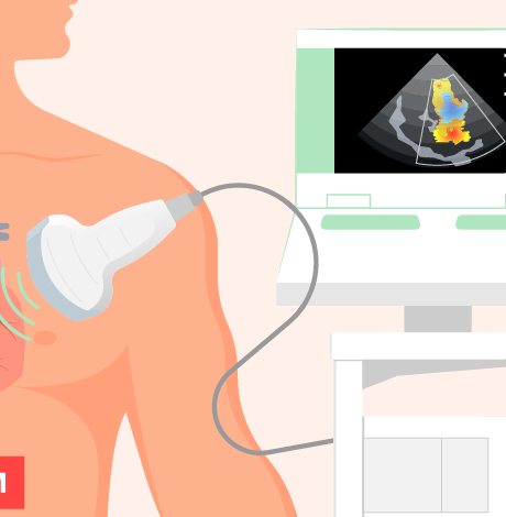
A 2D Echo (two-dimensional echocardiography) is a non-invasive heart imaging test that uses harmless ultrasound sound waves to create clear, real-time moving images of the heart. During the scan, a handheld probe is placed on the chest, which sends sound waves that bounce off the heart structures and return as detailed pictures on a screen. These images enable doctors to visualize the heart’s chambers, walls, valves, and major blood vessels as they move, allowing them to understand how the heart is functioning at that precise moment. The test does not involve radiation, injections, or pain, making it safe for people of all ages, including elderly patients and those who require repeated evaluations.
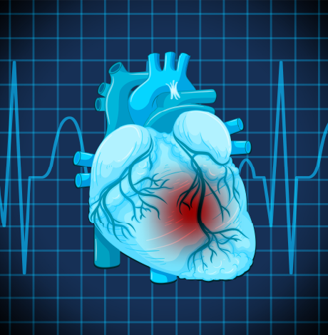
A 2D Echo plays a crucial role in diagnosing and monitoring a wide range of heart conditions. It helps assess the heart’s pumping strength, detect valve narrowing or leakage, identify abnormal thickening or weakening of the heart muscle, and reveal fluid collection around the heart. It is commonly recommended for symptoms such as breathlessness, chest discomfort, palpitations, leg swelling, or heart murmurs, as well as for routine follow-up in known cardiac patients. By providing accurate, visual information about heart structure and function, a 2D Echo supports timely diagnosis, effective treatment planning, and long-term cardiac care with clarity and confidence.


 Appointment
 Second Opinion
 Call
Call More
More


